
Page 314 - Libro 2
P. 314
294
PART 5 — ABDOMINAL
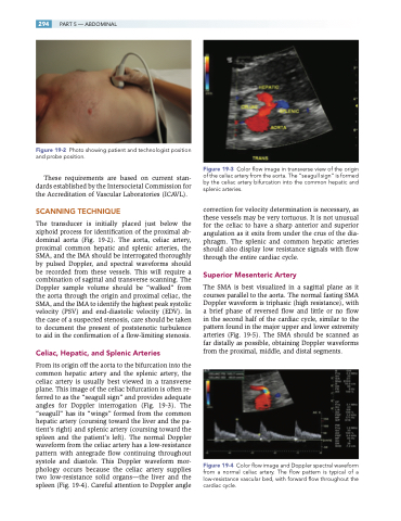
Figure 19-2 Photo showing patient and technologist position and probe position.
These requirements are based on current stan- dards established by the Intersocietal Commission for the Accreditation of Vascular Laboratories (ICAVL).
SCANNING TECHNIQUE
The transducer is initially placed just below the xiphoid process for identification of the proximal ab- dominal aorta (Fig. 19-2). The aorta, celiac artery, proximal common hepatic and splenic arteries, the SMA, and the IMA should be interrogated thoroughly by pulsed Doppler, and spectral waveforms should be recorded from these vessels. This will require a combination of sagittal and transverse scanning. The Doppler sample volume should be “walked” from the aorta through the origin and proximal celiac, the SMA, and the IMA to identify the highest peak systolic velocity (PSV) and end-diastolic velocity (EDV). In the case of a suspected stenosis, care should be taken to document the present of poststenotic turbulence to aid in the confirmation of a flow-limiting stenosis.
Celiac, Hepatic, and Splenic Arteries
From its origin off the aorta to the bifurcation into the common hepatic artery and the splenic artery, the celiac artery is usually best viewed in a transverse plane. This image of the celiac bifurcation is often re- ferred to as the “seagull sign” and provides adequate angles for Doppler interrogation (Fig. 19-3). The “seagull” has its “wings” formed from the common hepatic artery (coursing toward the liver and the pa- tient’s right) and splenic artery (coursing toward the spleen and the patient’s left). The normal Doppler waveform from the celiac artery has a low-resistance pattern with antegrade flow continuing throughout systole and diastole. This Doppler waveform mor- phology occurs because the celiac artery supplies two low-resistance solid organs—the liver and the spleen (Fig. 19-4). Careful attention to Doppler angle
Figure 19-3 Color flow image in transverse view of the origin of the celiac artery from the aorta. The “seagull sign” is formed by the celiac artery bifurcation into the common hepatic and splenic arteries.
correction for velocity determination is necessary, as these vessels may be very tortuous. It is not unusual for the celiac to have a sharp anterior and superior angulation as it exits from under the crus of the dia- phragm. The splenic and common hepatic arteries should also display low resistance signals with flow through the entire cardiac cycle.
Superior Mesenteric Artery
The SMA is best visualized in a sagittal plane as it courses parallel to the aorta. The normal fasting SMA Doppler waveform is triphasic (high resistance), with a brief phase of reversed flow and little or no flow in the second half of the cardiac cycle, similar to the pattern found in the major upper and lower extremity arteries (Fig. 19-5). The SMA should be scanned as far distally as possible, obtaining Doppler waveforms from the proximal, middle, and distal segments.
Figure 19-4 Color flow image and Doppler spectral waveform from a normal celiac artery. The flow pattern is typical of a low-resistance vascular bed, with forward flow throughout the cardiac cycle.
