
Page 321 - Libro 2
P. 321
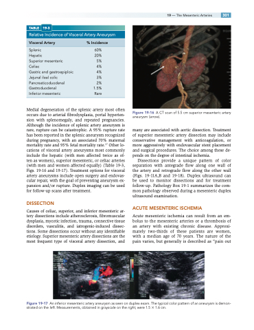
TABLE 19-3
Relative Incidence of Visceral Artery Aneurysm
Visceral Artery % Incidence
Splenic 60% Hepatic 20% Superior mesenteric 5% Celiac 4% Gastric and gastroepiploic 4% Jejunal ileal colic 3% Pancreaticoduodenal 2% Gastroduodenal 1.5% Inferior mesenteric Rare
Medial degeneration of the splenic artery most often occurs due to arterial fibrodysplasia, portal hyperten- sion with splenomegaly, and repeated pregnancies. Although the incidence of splenic artery aneurysm is rare, rupture can be catastrophic. A 95% rupture rate has been reported in the splenic aneurysm recognized during pregnancy, with an associated 70% maternal mortality rate and 95% fetal mortality rate.17 Other lo- cations of visceral artery aneurysms most commonly include the hepatic (with men affected twice as of- ten as women), superior mesenteric, or celiac arteries (with men and women affected equally) (Table 19-3, Figs. 19-16 and 19-17). Treatment options for visceral artery aneurysms include open surgery and endovas- cular repair, with the goal of preventing aneurysm ex- pansion and/or rupture. Duplex imaging can be used for follow-up scans after treatment.
DISSECTION
Causes of celiac, superior, and inferior mesenteric ar- tery dissections include atherosclerosis, fibromuscular dysplasia, mycotic infection, trauma, connective tissue disorders, vasculitis, and iatrogenic-induced dissec- tions. Some dissections occur without any identifiable etiology. Superior mesenteric artery dissections are the most frequent type of visceral artery dissection, and
Figure 19-16 A CT scan of 5.5 cm superior mesenteric artery aneurysm (arrow).
many are associated with aortic dissection. Treatment of superior mesenteric artery dissection may include conservative management with anticoagulation, or more aggressively with endovascular stent placement and surgical procedures. The choice among these de- pends on the degree of intestinal ischemia.
Dissections provide a unique pattern of color separation with antegrade flow along one wall of the artery and retrograde flow along the other wall (Figs. 19-11A,B and 19-18). Duplex ultrasound can be used to monitor dissections and for treatment follow-up. Pathology Box 19-1 summarizes the com- mon pathology observed during a mesenteric duplex ultrasound examination.
ACUTE MESENTERIC ISCHEMIA
Acute mesenteric ischemia can result from an em- bolus to the mesenteric arteries or a thrombosis of an artery with existing chronic disease. Approxi- mately two-thirds of these patients are women, with a median age of 70 years. The nature of the pain varies, but generally is described as “pain out
19 — The Mesenteric Arteries 301
Figure 19-17 An inferior mesenteric artery aneurysm as seen on duplex exam. The typical color pattern of an aneurysm is demon- strated on the left. Measurements, obtained in grayscale on the right, were 1.5 1.6 cm.
