
Page 338 - Libro 2
P. 338
318
PART 5 — ABDOMINAL
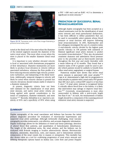
Figure 20-18 Transverse aortic color flow image illustrating a proximal renal artery stent
noted at the distal end of the stent when the diameter of the stented segment exceeds the diameter of the native renal artery. This most often occurs when the stent is placed in the smaller diameter distal renal artery.
It is important to note whether elevated velocity is focal or associated with downstream propagation of flow disturbance. Diameter mismatches are more likely to produce focal elevation in velocity without associated turbulence, whereas the flow profile of a flow-reducing stenosis exhibits high velocity, postste- notic turbulence, and dampening of the distal wave- form. Additionally, temporal changes in velocity will likely be noted when stenotic disease is present but velocity elevations should remain stable if related to a diameter mismatch.
At present, diagnostic criteria have not been well validated for the classification of renal artery stent stenosis, and native renal artery criteria are being applied with special consideration to the issues of compliance, diameter mismatch, and stent location.44–47 Recently, Mohabbat et al. reported a sen- sitivity of 93% and a specificity of 99% when using
SUMMARY
a PSV 280 cm/s and an RAR 4.5 to determine a significant in-stent stenosis.46
PREDICTION OF SUCCESSFUL RENAL REVASCULARIZATION
Although duplex sonography has been accepted as a valued noninvasive tool for the identification of renal artery stenosis and renal parenchymal dysfunction, it has been difficult to define parameters that can be used to successfully select patients whose blood pressure or renal function will improve following renal revascularization. In 2001, Radermacher and his colleagues investigated the use of a resistive index (1-end-diastolic velocity divided by the highest peak systolic velocity) in 138 patients with unilateral or bilateral significant renal artery stenosis to predict successful revascularization.48 They also documented creatinine clearance and ambulatory blood pressure prior to the procedure and at three-month intervals throughout the first year and yearly thereafter with a mean follow-up of 32 months. They found that a resistive index of 80 or greater could be used to pre- dict which patients would be unlikely to benefit from either renal artery angioplasty or surgical bypass.
Numerous studies have shown that severe renal artery stenosis is associated with renal atrophy.49–54 Caps et al. demonstrated a high risk for progression to renal atrophy in patients with a renal artery PSV greater than 400 cm/s and a cortical EDV equal to or less than 5 cm/s.55 If renal artery stenosis is recognized before it critically affects blood flow to the kidney, it is likely that intervention may salvage or improve renal func- tion.54,56 Conversely, revascularization is most often unsuccessful in kidneys with a pole-to-pole length less than 8 cm.57 As such, the measurement of kidney length should be a component of the scanning protocol whenever renal artery stenosis is suspected.
Duplex sonography of the renal vasculature and kidneys has become the initial primary diagnostic procedure for evaluation of renovascular hypertension and suspected renal artery pathology. Although technically challenging, renal vascular sonography provides noninvasive, cost-effective, and accurate diagnostic information. Duplex technology allows for the definition of vessel and tissue pathology, detection of altered blood flow patterns, and quantitation of hemodynamic disturbances. The length of the abdominal aorta, the renal arteries, and the kidneys can be evaluated with B-mode imaging to localize atherosclerotic disease, fibromuscular dysplasia, aneurysms, dissections, cysts, and masses, and to demonstrate extrinsic compression of the renal vessels due to masses or entrapment. This modality has also shown value as a tool for confirming progression of renal artery stenosis and defining therapeutic options based on kidney size. Color flow and power Doppler
