
Page 345 - Libro 2
P. 345
21 — The Inferior Vena Cava and Iliac Veins
325
disperse bowel gas and obtain better images of deep abdominal structures. The amount of force exerted on the transducer must be regulated because exces- sive pressure will also compress the IVC, making it difficult to visualize.
SCANNING TECHNIQUE
Achieving adequate penetration usually requires a 1- to 4-MHz probe; however, a 5-MHz probe may be more appropriate for thin patients. A complete exam requires a longitudinal and transverse survey of the IVC from the diaphragm to the confluence of the common iliac veins. The exam begins with the probe perpendicular, at the midline of the body, and just distal to the xiphoid process of the sternum. Angling the transducer to the patient’s left allows visualiza- tion of the proximal abdominal aorta posterior to the liver. After identifying this landmark, the transducer can be angled to the patient’s right side to obtain a longitudinal image of the IVC posterior to the liver (Fig. 21-1). The hepatic veins, which are anterior tributaries of the proximal IVC, should be recognized and evaluated. The transducer can then be slowly moved inferiorly using a rock-and-slide motion. By slightly rocking to the right and then to the left, each side of the IVC is scanned while sliding the probe inferiorly.5 Rotating the transducer may be required to keep the IVC in view. This technique uses the sag- ittal plane to obtain longitudinal images of the proxi- mal, mid, and distal IVC to the common iliac vein confluence (usually at the level of the umbilicus).
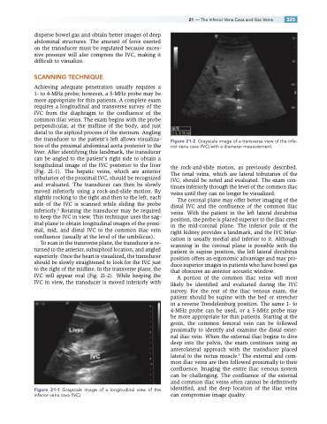
To scan in the transverse plane, the transducer is re- turned to the anterior, subxiphoid location, and angled superiorly. Once the heart is visualized, the transducer should be slowly straightened to look for the IVC just to the right of the midline. In the transverse plane, the IVC will appear oval (Fig. 21-2). While keeping the IVC in view, the transducer is moved inferiorly with
Figure 21-1 Grayscale image of a longitudinal view of the inferior vena cava (IVC).
Figure 21-2 Grayscale image of a transverse view of the infe- rior vena cava (IVC) with a diameter measurement.
the rock-and-slide motion, as previously described. The renal veins, which are lateral tributaries of the IVC, should be noted and evaluated. The exam con- tinues inferiorly through the level of the common iliac veins until they can no longer be visualized.
The coronal plane may offer better imaging of the distal IVC and the confluence of the common iliac veins. With the patient in the left lateral decubitus position, the probe is placed superior to the iliac crest in the mid-coronal plane. The inferior pole of the right kidney provides a landmark, and the IVC bifur- cation is usually medial and inferior to it. Although scanning in the coronal plane is possible with the patient in supine position, the left lateral decubitus position offers an ergonomic advantage and may pro- duce superior images in patients who have bowel gas that obscures an anterior acoustic window.
A portion of the common iliac veins will most likely be identified and evaluated during the IVC survey. For the rest of the iliac venous exam, the patient should be supine with the bed or stretcher in a reverse Trendelenburg position. The same 1- to 4-MHz probe can be used, or a 5-MHz probe may be more appropriate for thin patients. Starting at the groin, the common femoral vein can be followed proximally to identify and examine the distal exter- nal iliac vein. When the external iliac begins to dive deep into the pelvis, the exam continues using an anterolateral approach with the transducer placed lateral to the rectus muscle.6 The external and com- mon iliac veins are then followed proximally to their confluence. Imaging the entire iliac venous system can be challenging. The confluence of the external and common iliac veins often cannot be definitively identified, and the deep location of the iliac veins can compromise image quality.
