
Page 346 - Libro 2
P. 346
326 PART 5 — ABDOMINAL
TABLE 21-2
Required Images for IVC and Iliac Veins
Image Plane
Longitudinal
Transverse
Anatomic Level
Proximal IVC
Middle IVC
Distal IVC
Common iliac vein confluence External iliac veins
Proximal IVC
Middle IVC
Distal IVC
Common iliac vein confluence External iliac veins
Landmarks
Diaphragm and hepatic vein(s) Head of the pancreas
Common iliac veins
Hepatic veins Renal veins
Common iliac veins
changes in abdominal pressure produced during res- piration. The grayscale image can be evaluated for various pathology including thrombosis, intralumi- nal tumors, and extrinsic compressions.
THROMBOSIS
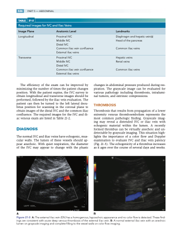
Thrombosis that results from propagation of a lower extremity venous thromboembolism represents the most common pathologic finding. Grayscale imag- ing may reveal a distended IVC or iliac vein with echogenic material within the lumen. A recently formed thrombus can be virtually anechoic and un- detectable by grayscale imaging. This situation high- lights the importance of a color flow and Doppler examination to evaluate IVC and iliac vein patency (Fig. 21-3). The echogenicity of a thrombus increases as it ages over the course of several days and weeks.
The efficiency of the exam can be improved by minimizing the number of times the patient changes position. With the patient supine, the IVC survey to obtain longitudinal and transverse images should be performed, followed by the iliac vein evaluation. The patient can then be turned to the left lateral decu- bitus position for scanning in the coronal plane to obtain images of the distal IVC and the common iliac confluence. The required images for the IVC and ili- ac venous exam are listed in Table 21-2.
DIAGNOSIS
The normal IVC and iliac veins have echogenic, mus- cular walls. The lumen of these vessels should ap- pear anechoic. With quiet respiration, the diameter of the IVC may appear to change with the phasic
AB
Figure 21-3 A: The external iliac vein (EIV ) has a homogenous, hypoechoic appearance and no color flow is detected. These find- ings are consistent with acute deep venous thrombosis of the external iliac vein. B: A normal external iliac vein with an anechoic lumen on grayscale imaging and complete filling to the vessel walls on color flow imaging.
