
Page 348 - Libro 2
P. 348
328
PART 5 — ABDOMINAL
the fistula. Directly imaging the connection between the aorta and the IVC may be difficult. Surgically cre- ated portacaval fistulas usually involve a side-to-side connection between the portal vein and the IVC. The fistula creates a tissue bruit, and optimal visualization often requires using the liver as an acoustic window with the patient turned onto his or her left side.
The deep location and relatively low flow state of the IVC and iliac veins stress the limits of the color Doppler exam. Power Doppler offers a complemen- tary assessment that can overcome these imaging challenges. Because power Doppler functions inde- pendent of the ultrasound angle of incidence, it can evaluate patency even when the only images obtain- able are in the transverse plane. Power Doppler can also detect slow flow better than color Doppler. This increased sensitivity allows power Doppler to detect extremely low flow in the IVC or iliac veins, which could otherwise result in a false-positive diagnosis of venous thrombosis based on the color Doppler findings alone. The use of power Doppler to detect and define low flow conditions such as intrahepatic portacaval shunts has also been reported.10
SPECTRAL DOPPLER CHARACTERISTICS
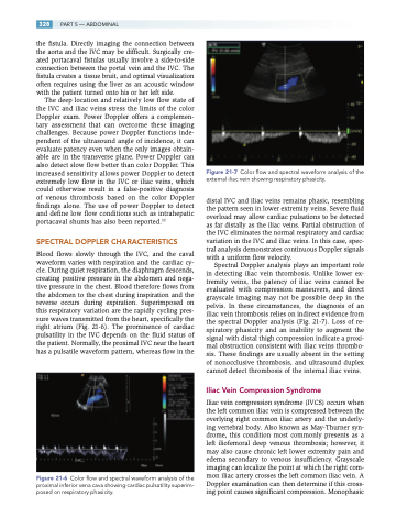
Blood flows slowly through the IVC, and the caval waveform varies with respiration and the cardiac cy- cle. During quiet respiration, the diaphragm descends, creating positive pressure in the abdomen and nega- tive pressure in the chest. Blood therefore flows from the abdomen to the chest during inspiration and the reverse occurs during expiration. Superimposed on this respiratory variation are the rapidly cycling pres- sure waves transmitted from the heart, specifically the right atrium (Fig. 21-6). The prominence of cardiac pulsatility in the IVC depends on the fluid status of the patient. Normally, the proximal IVC near the heart has a pulsatile waveform pattern, whereas flow in the
Figure 21-6 Color flow and spectral waveform analysis of the proximal inferior vena cava showing cardiac pulsatility superim- posed on respiratory phasicity.
Figure 21-7 Color flow and spectral waveform analysis of the external iliac vein showing respiratory phasicity.
distal IVC and iliac veins remains phasic, resembling the pattern seen in lower extremity veins. Severe fluid overload may allow cardiac pulsations to be detected as far distally as the iliac veins. Partial obstruction of the IVC eliminates the normal respiratory and cardiac variation in the IVC and iliac veins. In this case, spec- tral analysis demonstrates continuous Doppler signals with a uniform flow velocity.
Spectral Doppler analysis plays an important role in detecting iliac vein thrombosis. Unlike lower ex- tremity veins, the patency of iliac veins cannot be evaluated with compression maneuvers, and direct grayscale imaging may not be possible deep in the pelvis. In these circumstances, the diagnosis of an iliac vein thrombosis relies on indirect evidence from the spectral Doppler analysis (Fig. 21-7). Loss of re- spiratory phasicity and an inability to augment the signal with distal thigh compression indicate a proxi- mal obstruction consistent with iliac veins thrombo- sis. These findings are usually absent in the setting of nonocclusive thrombosis, and ultrasound duplex cannot detect thrombosis of the internal iliac veins.
Iliac Vein Compression Syndrome
Iliac vein compression syndrome (IVCS) occurs when the left common iliac vein is compressed between the overlying right common iliac artery and the underly- ing vertebral body. Also known as May-Thurner syn- drome, this condition most commonly presents as a left iliofemoral deep venous thrombosis; however, it may also cause chronic left lower extremity pain and edema secondary to venous insufficiency. Grayscale imaging can localize the point at which the right com- mon iliac artery crosses the left common iliac vein. A Doppler examination can then determine if this cross- ing point causes significant compression. Monophasic
