
Page 349 - Libro 2
P. 349
21 — The Inferior Vena Cava and Iliac Veins
329
flow without respiratory variation distal to the com- pression and increased flow velocity at the point of compression are signs of a localized iliac vein steno- sis consistent with IVCS.11 Definitive imaging studies including venography with intraluminal pressures, intravascular ultrasonography, CT, or magnetic reso- nance imaging may be required to confirm the diag- nosis. Pathology Box 21-1 lists the common pathology encountered during the ultrasound examination of the inferior vena cava and the iliac veins.
DUPLEX ULTRASOUND GUIDANCE FOR IVC FILTER PLACEMENT
The utility of duplex ultrasound has expanded be- yond the diagnosis of a thrombus to now include guidance during IVC filter placement. Percutaneous IVC filter placement has traditionally been performed with contrast venography in an operating room or interventional radiology suite. Duplex ultrasound guidance can transform IVC filter placement into a bedside procedure that does not require exposure to radiation or intravenous contrast.12
PATIENT PREPARATION AND POSITIONING
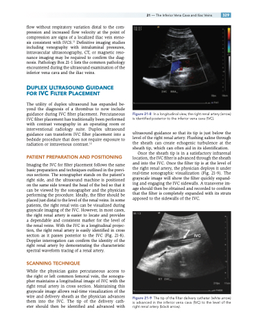
Imaging the IVC for filter placement follows the same basic preparation and techniques outlined in the previ- ous sections. The sonographer stands on the patient’s right side, and the ultrasound machine is positioned on the same side toward the head of the bed so that it can be viewed by the sonographer and the physician performing the procedure. Ideally, the filter should be placed just distal to the level of the renal veins. In some patients, the right renal vein can be visualized during grayscale imaging of the IVC. However, in most cases, the right renal artery is easier to locate and provides a dependable and consistent marker for the level of the renal veins. With the IVC in a longitudinal projec- tion, the right renal artery is easily identified in cross section as it passes posterior to the IVC (Fig. 21-8). Doppler interrogation can confirm the identity of the right renal artery by demonstrating the characteristic spectral waveform tracing of a renal artery.
SCANNING TECHNIQUE
While the physician gains percutaneous access to the right or left common femoral vein, the sonogra- pher maintains a longitudinal image of IVC with the right renal artery in cross section. Maintaining this grayscale image allows real-time visualization of the wire and delivery sheath as the physician advances them into the IVC. The tip of the delivery cath- eter should then be identified and advanced with
In a longitudinal view, the right renal artery (arrow) is identified posterior to the inferior vena cava (IVC).
ultrasound guidance so that its tip is just below the level of the right renal artery. Flushing saline through the sheath can create echogenic turbulence at the sheath tip, which can often aid in its identification.
Once the sheath tip is in a satisfactory infrarenal location, the IVC filter is advanced through the sheath and into the IVC. Once the filter tip is at the level of the right renal artery, the physician deploys it under real-time sonographic visualization (Fig. 21-9). The grayscale image will show the filter quickly expand- ing and engaging the IVC sidewalls. A transverse im- age should then be obtained and recorded to confirm that the filter is completely expanded with its struts apposed to the sidewalls of the IVC.
Figure 21-9 The tip of the filter delivery catheter (white arrow) is advanced in the inferior vena cava (IVC) to the level of the right renal artery (black arrow).
Figure 21-8
