
Page 347 - Libro 2
P. 347
21 — The Inferior Vena Cava and Iliac Veins
327
A thrombus that does not obstruct flow may only be detected by grayscale imaging demonstrating free floating echogenic material within the IVC or iliac vein lumen.
NEOPLASTIC OBSTRUCTION
Compared to thrombotic occlusion, neoplastic ob- struction of the IVC and iliac veins is rare. Grayscale imaging reveals an intraluminal tumor or an extrin- sic mass. Intraluminal tumors typically arise from hepatic or renal veins and may secondarily obstruct or thrombose the IVC.7 Extrinsic tumors may com- pletely or partially obstruct the IVC or iliac veins, resulting in dilated collateral veins and distention of the distal IVC and iliac veins. An intraluminal tumor is typically moderately echogenic and will demon- strate flow within the mass on color flow imaging. Small vessels can be seen within the tumor itself.
INFERIOR VENA CAVAL INTERRUPTION
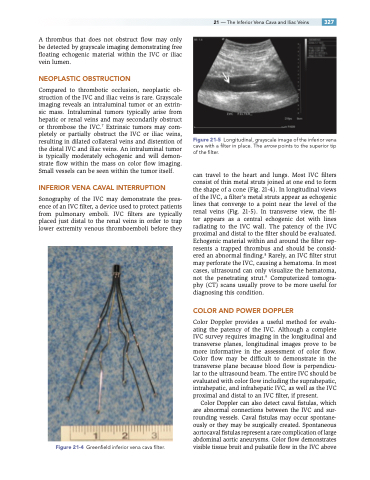
Sonography of the IVC may demonstrate the pres- ence of an IVC filter, a device used to protect patients from pulmonary emboli. IVC filters are typically placed just distal to the renal veins in order to trap lower extremity venous thromboemboli before they
Figure 21-4 Greenfield inferior vena cava filter.
Figure 21-5 Longitudinal, grayscale image of the inferior vena cava with a filter in place. The arrow points to the superior tip of the filter.
can travel to the heart and lungs. Most IVC filters consist of thin metal struts joined at one end to form the shape of a cone (Fig. 21-4). In longitudinal views of the IVC, a filter’s metal struts appear as echogenic lines that converge to a point near the level of the renal veins (Fig. 21-5). In transverse view, the fil- ter appears as a central echogenic dot with lines radiating to the IVC wall. The patency of the IVC proximal and distal to the filter should be evaluated. Echogenic material within and around the filter rep- resents a trapped thrombus and should be consid- ered an abnormal finding.8 Rarely, an IVC filter strut may perforate the IVC, causing a hematoma. In most cases, ultrasound can only visualize the hematoma, not the penetrating strut.9 Computerized tomogra- phy (CT) scans usually prove to be more useful for diagnosing this condition.
COLOR AND POWER DOPPLER
Color Doppler provides a useful method for evalu- ating the patency of the IVC. Although a complete IVC survey requires imaging in the longitudinal and transverse planes, longitudinal images prove to be more informative in the assessment of color flow. Color flow may be difficult to demonstrate in the transverse plane because blood flow is perpendicu- lar to the ultrasound beam. The entire IVC should be evaluated with color flow including the suprahepatic, intrahepatic, and infrahepatic IVC, as well as the IVC proximal and distal to an IVC filter, if present.
Color Doppler can also detect caval fistulas, which are abnormal connections between the IVC and sur- rounding vessels. Caval fistulas may occur spontane- ously or they may be surgically created. Spontaneous aortocaval fistulas represent a rare complication of large abdominal aortic aneurysms. Color flow demonstrates visible tissue bruit and pulsatile flow in the IVC above
