
Page 359 - Libro 2
P. 359
22 — The Hepatoportal System 339
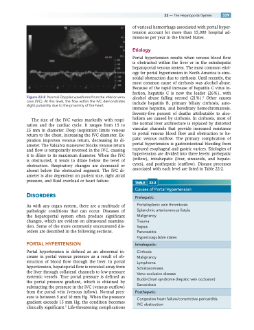
Normal Doppler waveforms from the inferior vena cava (IVC). At this level, the flow within the IVC demonstrates slight pulsatility due to the proximity of the heart.
The size of the IVC varies markedly with respi- ration and the cardiac cycle. It ranges from 15 to 25 mm in diameter. Deep inspiration limits venous return to the chest, increasing the IVC diameter. Ex- piration improves venous return, decreasing its di- ameter. The Valsalva maneuver blocks venous return and flow is temporarily reversed in the IVC, causing it to dilate to its maximum diameter. When the IVC is obstructed, it tends to dilate below the level of obstruction. Respiratory changes are decreased or absent below the obstructed segment. The IVC di- ameter is also dependent on patient size, right atrial pressure, and fluid overload or heart failure.
DISORDERS
As with any organ system, there are a multitude of pathologic conditions that can occur. Diseases of the hepatoportal system often produce significant changes, which are evident on ultrasound examina- tion. Some of the more commonly encountered dis- orders are described in the following sections.
PORTAL HYPERTENSION
Portal hypertension is defined as an abnormal in- crease in portal venous pressure as a result of ob- struction of blood flow through the liver. In portal hypertension, hepatopetal flow is rerouted away from the liver through collateral channels to low-pressure systemic vessels. True portal pressure is defined as the portal pressure gradient, which is obtained by subtracting the pressure in the IVC (venous outflow) from the portal vein (venous inflow). Normal pres- sure is between 5 and 10 mm Hg. When the pressure gradient exceeds 15 mm Hg, the condition becomes clinically significant.2 Life-threatening complications
of variceal hemorrhage associated with portal hyper- tension account for more than 15,000 hospital ad- missions per year in the United States.
Etiology
Portal hypertension results when venous blood flow is obstructed within the liver or in the extrahepatic hepatoportal venous system. The most common etiol- ogy for portal hypertension in North America is sinu- soidal obstruction due to cirrhosis. Until recently, the most common cause of cirrhosis was alcohol abuse. Because of the rapid increase of hepatitis C virus in- fection, hepatitis C is now the leader (26%), with alcohol abuse falling second (21%).9 Other causes include hepatitis B, primary biliary cirrhosis, auto- immune hepatitis, and hereditary hemochromatosis. Seventy-five percent of deaths attributable to alco- holism are caused by cirrhosis. In cirrhosis, most of the normal liver architecture is replaced by distorted vascular channels that provide increased resistance to portal venous blood flow and obstruction to he- patic venous outflow. The primary complication of portal hypertension is gastrointestinal bleeding from ruptured esophageal and gastric varices. Etiologies of hypertension are divided into three levels: prehepatic (inflow), intrahepatic (liver, sinusoids, and hepato- cytes), and posthepatic (outflow). Disease processes associated with each level are listed in Table 22-2.
TABLE 22-2
Causes of Portal Hypertension
Prehepatic:
Portal/splenic vein thrombosis Splanchnic arteriovenous fistula Malignancy
Trauma
Sepsis
Pancreatitis Hypercoagulable states
Intrahepatic:
Cirrhosis
Malignancy
Lymphoma
Schistosomiasis
Veno-occlusive disease
Budd-Chiari syndrome (hepatic vein occlusion) Sarcoidosis
Posthepatic:
Congestive heart failure/constrictive pericarditis IVC obstruction
Figure 22-8
