
Page 361 - Libro 2
P. 361
portosystemic collaterals. In the splenic hilum, di- lated veins sometimes show anastomoses to veins of the stomach or esophagus.
Portosystemic Collateral Anatomy
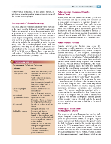
Detection of portosystemic collateral veins (varices) is the most specific finding of portal hypertension. Varices are reported to occur in approximately 39% of patients with biopsy-proven cirrhosis and are more common in patients with advanced disease.13 Color duplex sonography visualizes approximately 65% to 90% of collateral shunts.14 Commonly seen collaterals include the paraumbilical vein, the cor- onary vein, the gastroesophageal veins, and the splenorenal vein (Fig. 22-11). The most common col- lateral shunt is the coronary-gastroesophageal route (80% to 90%); when dilated, these cause esopha- geal varices.14 Pathology Box 22-2 describes various portosystemic collateral pathways.2,10
PATHOLOGY BOX 22-2
22 — The Hepatoportal System 341 Arterialization (Increased Hepatic
Artery Flow)
When portal venous pressure increases, portal vein flow decreases and hepatic artery flow increases as a homeostatic mechanism to maintain hepatic per- fusion. Enlargement, increased flow, and a tortuous “corkscrew” appearance are commonly seen in pa- tients with cirrhosis, portal hypertension, portal vein thrombosis, and inflammation related to chronic ac- tive hepatitis. Color duplex imaging demonstrates an enlarged hepatic artery with high velocity turbulent flow (mixed color flow) referred to as “arterialization.”
Arteriovenous Fistulae
Hepatic arterial–portal fistulae may cause life- threatening portal hypertension. Causes of arterial– portal fistulae include penetrating trauma, iatrogenic trauma secondary to liver biopsies, transhepatic cholangiography, and transhepatic catheterization of the bile ducts or portal veins. Profuse collateral flow typically accompanies severe portal hypertension in patients with hepatic artery to portal vein commu- nications. With an arterial–portal fistula, the result- ing pressure gradient causes blood to flow from the artery to the vein. Duplex sonography demonstrates arterialized hepatofugal flow in the portal vein. Large anechoic spaces are seen within the liver in the area of the communication. Color Doppler shows a tur- bulent high-velocity flow “color bruit” detected be- tween the communication of the artery and the vein.
Another type of fistula can arise between the por- tal vein and a hepatic vein. The causes responsible for venovenous fistulae are trauma, surgery, organ punctures, perforated aneurysms, and idiopathic causes. The pressure gradients associated with ve- novenous fistulae cause blood to flow from the por- tal vein to the hepatic vein. In cases of hepatic vein to portal vein fistulae, the triphasic spectral pattern of the hepatic veins is reflected back to the portal system, producing increased pulsatility in the portal vein waveform.
Transjugular Intrahepatic Portosystemic Shunts
The treatment of portal hypertension involves tech- niques designed to decrease the pressure or decom- press the portal venous system. In the past, surgical shunts were created that connected the portal vein to the IVC (portocaval shunt), the superior mesenteric vein to the IVC (mesocaval shunt), or the splenic vein to the left renal vein (splenorenal shunt). These all reduced flow into the liver and thus helped lessen the pressure in the portal venous system. Current therapy involves a percutaneously created shunt, which avoids
Portosystemic Collateral Pathways
Collateral Features
Coronary vein
(left gastric vein)
Gastroesophageal veins
Recanalized paraumbilical vein
Splenorenal
Gallbladder varices
• Most often joins the portal system at the splenoportal confluence
• With portal hypertension, diameter 7 mm
• Can demonstrate hepatofugal flow, which often leads to esophageal varices
• Located posterior to left lobe of liver
• Can be large and tortuous
• Located in the fissure for the
ligamentum teres
• Diameter 3 mm is diagnos-
tic for portal hypertension
• Prominent varices at splenic
hilum
• Enlarged left renal vein • Portosystemic shunts
between the cystic vein branch of the portal vein and either anterior abdominal wall veins or portal vein branches within the liver
• Found in 30% of patients with portal hypertension
• 3–8 mm in diameter
• Tortuous vessels along gallbladder wall
