
Page 366 - Libro 2
P. 366
346 PART 5 — ABDOMINAL
by creating hepatic vein congestion. Obstruction of the IVC may be caused by congenital stenosis or oc- clusion, thrombosis from hypercoagulable states, or tumor invasion.
Hepatic venous obstruction is often accompanied by stenosis or obstruction of the IVC. When the IVC is involved, lower extremity edema maybe present. With severe obstruction, collateral channels are formed as follows: portal vein collaterals via en- larged paraumbilical veins, reversed hepatic venous flow to systemic or capsular veins, and intrahepatic venous to hepatic venous collaterals. Flow in the portal vein maybe slow or reversed. The caudate lobe enlarges due to blood volume overload as the other lobes of the liver will try to drain out via the caudate veins, which empty directly into the IVC.4 The sonographic findings of Budd-Chiari syndrome are listed in Pathology Box 22-5.
SUMMARY
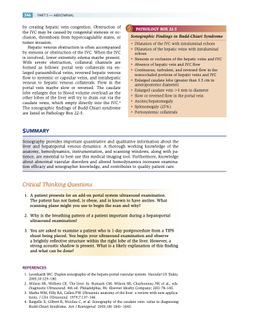
PATHOLOGY BOX 22-5
Sonographic Findings in Budd-Chiari Syndrome
• Dilatation of the IVC with intraluminal echoes
• Dilatation of the hepatic veins with intraluminal
echoes
• Stenosis or occlusion of the hepatic veins and IVC
• Absence of hepatic vein and IVC flow
• Continuous, turbulent, and reversed flow in the
nonoccluded portions of hepatic veins and IVC
• Enlarged caudate lobe (greater than 3.5 cm in
anteroposterior diameter)
• Enlarged caudate vein 3 mm in diameter
• Slow or reversed flow in the portal vein
• Ascites/hepatomegaly
• Splenomegaly (25%)
• Portosystemic collaterals
Sonography provides important quantitative and qualitative information about the liver and hepatoportal venous dynamics. A thorough working knowledge of the anatomy, hemodynamics, instrumentation, and scanning windows, along with pa- tience, are essential to best use this medical imaging tool. Furthermore, knowledge about abnormal vascular disorders and altered hemodynamics increases examina- tion efficacy and sonographer knowledge, and contributes to quality patient care.
Critical Thinking Questions
1. A patient presents for an add-on portal system ultrasound examination. The patient has not fasted, is obese, and is known to have ascites. What scanning plane might you use to begin the scan and why?
2. Why is the breathing pattern of a patient important during a hepatoportal ultrasound examination?
3. You are asked to examine a patient who is 1-day postprocedure from a TIPS shunt being placed. You begin your ultrasound examination and observe
a brightly reflective structure within the right lobe of the liver. However, a strong acoustic shadow is present. What is a likely explanation of this finding and what can be done?
REFERENCES
1. Leonhardt WC. Duplex sonography of the hepato-portal vascular system. Vascular US Today. 2005;10:129–180.
2. Wilson SR, Withers CE. The liver. In: Rumack CM, Wilson SR, Charboneau JW, et al., eds. Diagnostic Ultrasound. 4th ed. Philadelphia, PA: Elsevier Mosby Company; 2011:78–145.
3. Marks WM, Filly RA, Callen PW. Ultrasonic anatomy of the liver: a review with new applica-
tions. J Clin Ultrasound. 1979;7:137–146.
4. Bargallo X, Gilbert R, Nicolau C, et al. Sonography of the caudate vein: value in diagnosing
Budd-Chiari Syndrome. Am J Roentgenol. 2003;181:1641–1645.
