
Page 364 - Libro 2
P. 364
344
PART 5 — ABDOMINAL
Figure 22-14 An image from a TIPS with a stenosis. Note the elevated velocity of 280 cm/s.
Doppler and color frequencies, color priority set- tings, and Doppler and color scale or pulse repetition frequencies.
PORTAL VENOUS THROMBOSIS
Thrombosis within the portal, splenic, and superior mesenteric veins can result from flow stasis secondary to cirrhosis and subsequent portal hypertension. As portal venous flow to the liver decreases, arterial flow increases. This is a homeostatic mechanism to main- tain hepatic perfusion. Other etiologies of portal vein thrombosis (PVT) include inflammatory processes (such as pancreatitis, appendicitis, and diverticulitis), various hypercoagulable states (including protein C or S deficiencies, antithrombin deficiency, and poly- cythemiavera),surgicalintervention,abdominalma- lignancy (hepatocellular or pancreatic carcinomas), sepsis, and trauma.1 Patients with hepatocellular
Figure 22-15 An occluded TIPS with no filling on color flow imaging and echogenic material within the shunt itself.
carcinoma (HCC) are at greater risk of developing ma- lignant thrombus (intravascular tumor) due to direct invasion of the portal vein. Pancreatitis and pancre- atic carcinoma are frequent causes of thrombosis and tumor infiltration in the portal, splenic, and superior mesenteric veins. A sudden onset of ascites, acute ab- dominal pain, and elevated D-dimer in patients may indicate the presence of PVT.
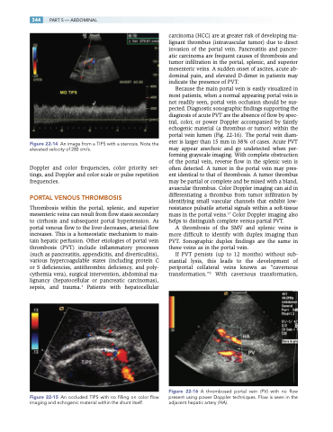
Because the main portal vein is easily visualized in most patients, when a normal appearing portal vein is not readily seen, portal vein occlusion should be sus- pected. Diagnostic sonographic findings supporting the diagnosis of acute PVT are the absence of flow by spec- tral, color, or power Doppler accompanied by faintly echogenic material (a thrombus or tumor) within the portal vein lumen (Fig. 22-16). The portal vein diam- eter is larger than 15 mm in 38% of cases. Acute PVT may appear anechoic and go undetected when per- forming grayscale imaging. With complete obstruction of the portal vein, reverse flow in the splenic vein is often detected. A tumor in the portal vein may pres- ent identical to that of thrombosis. A tumor thrombus may be partial or complete and be mixed with a bland, avascular thrombus. Color Doppler imaging can aid in differentiating a thrombus from tumor infiltration by identifying small vascular channels that exhibit low- resistance pulsatile arterial signals within a soft-tissue mass in the portal veins.17 Color Doppler imaging also helps to distinguish complete versus partial PVT.
A thrombosis of the SMV and splenic veins is more difficult to identify with duplex imaging than PVT. Sonographic duplex findings are the same in these veins as in the portal vein.
If PVT persists (up to 12 months) without sub- stantial lysis, this leads to the development of periportal collateral veins known as “cavernous transformation.”2 With cavernous transformation,
Figure 22-16 A thrombosed portal vein (PV ) with no flow present using power Doppler techniques. Flow is seen in the adjacent hepatic artery (HA).
