
Page 365 - Libro 2
P. 365
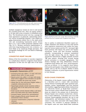
Figure 22-17 A thrombosed portal vein with cavernous trans- formation (arrows indicate the periportal collateral veins).
multiple serpiginous vessels are seen in and around the occluded portal vein. They can appear within 6 to 20 days after acute occlusion to reestablish portal flow. The thrombosed portal vein has a sponge-like mass appearance on grayscale, representing numer- ous venous recanalizing channels. Color duplex im- aging demonstrates absent flow in the main portal vein and recanalizing hepatopetal portal venous flow (2 to 7 cm/s) within periportal collateral veins (Fig. 22-17). Because cavernous transformation re- sults from long-standing portal vein occlusion, it is more likely to be caused by benign processes. The sonographic findings for PVT are listed in Pathology Box 22-4.
CONGESTIVE HEART FAILURE
Edema of the liver secondary to vascular congestion is a complication related to congestive heart failure. Impedance of flow into the right side of the heart
PATHOLOGY BOX 22-4
Figure 22-18 A portal vein image demonstrating markedly pulsatile flow from a patient with congestive heart failure.
due to cardiac or pulmonary disorders causes sec- ondary dilatation and absence of vein wall motion with respiratory maneuvers both within the hepa- toportal venous system and the IVC. Increased right heart pressure will impact the portal and hepatic waveforms. Portal vein flow becomes markedly pulsatile, corresponding to pressure transmitted from the right atrium (Fig. 22-18). Hepatic vein waveforms demonstrate a highly pulsatile inverted “W”-type pattern, showing flow reversal during systole secondary to tricuspid regurgitation. Two distinct findings that help differentiate between congestive heart failure and “true” portal hyperten- sion are as follows: (1) In congestive heart failure, both portal and hepatic veins exhibit increased pul- satility, and (2) the IVC is dilated. Neither of these findings is indicative of portal hypertension related to liver disease.
BUDD-CHIARI SYNDROME
Obstruction of the hepatic venous outflow tract due to a thrombus and or a tumor with concomitant clinical features including right upper quadrant pain, jaundice, ascites, hepatomegaly, and liver function abnormalities suggesting hepatocellular dysfunctions are collectively known as Budd-Chiari syndrome.1 There are many causes of Budd-Chiari syndrome, and these are related to the primary site of obstruction. Primary hepatic vein occlusion results from a throm- bus or tumor infiltration. A thrombus can occur with cirrhosis, hypercoagulable disorders, the use of oral contraceptives, and abdominal trauma.10 Tumor in- vasion is most often associated with hepatocellular carcinoma. IVC occlusion or stenosis cephalad to the hepatic veins can cause Budd-Chiari syndrome
22 — The Hepatoportal System 345
Sonographic Characteristics of PVT
• Increased portal vein caliber (15 mm) with intra- luminal echoes (38%) of cases (acute)
• Massive portal vein caliber (23 mm) with intralu- minal echoes suggests tumor thrombus not specific
• Failure to visualize the portal vein (acute)
• Absent flow by color and power Doppler within a
completely obstructed portal vein
• Hepatofugal pulsatile arterial waveform within a
soft tissue mass in the main portal vein (tumor
thrombus)
• Small vascular arterial channels within a soft tissue
mass in the main portal vein (tumor thrombus)
• Increased hepatic arterial flow (arterialization)
• Small echogenic/fibrotic portal channels with
intraluminal echoes (chronic)
• Cavernous transformation; periportal collaterals
(chronic)
• Gallbladder varices
