
Page 376 - Libro 2
P. 376
356
PART 5 — ABDOMINAL
Figure 23-11 A postoperative perinephric hematoma (same patient as in Fig. 23-10). A color Doppler image demonstrates decreased cortical perfusion due to pressure from the sur- rounding fluid hematoma.
Urinomas form when urine leaks from either the ureteral anastomosis or a focal area of ureteral necrosis. These are usually discovered in the first few weeks posttransplant. Clinically, suspicion is raised when urine output decreases, especially in the absence of renal failure, or if there is leakage of urine from the surgical incision. Ultrasound will demonstrate a fluid collection, usually lo- cated between the kidney and the bladder. Urino- mas are typically anechoic unless superinfection has occurred, but some may contain septations (Fig. 23-13).
Lymphoceles occur when there is surgical disruption of the lymphatic chain. These col- lections usually appear 4 to 8 weeks postopera- tively. Typically, these collections are discovered incidentally. However, lymphoceles can compress the ureter causing obstruction of the collecting system or become superinfected, both of which require percutaneous drainage or surgical marsu- pialization. On US, lymphoceles are well-defined, anechoic fluid collections, which may demonstrate multiple thin septations (Fig. 23-14). It is impor- tant not to confuse a urinoma with a lymphocele. Remember that urinomas will occur within the first few weeks posttransplant, whereas lympho- celes will be seen in a later time frame, usually after the first month.
HYDRONEPHROSIS
Mild pelvicaliectasis (hydronephrosis) is a nor- mal finding postrenal transplantation because the denervated kidney loses its autonomic tone, allowing the intrarenal collecting system to di- late. Patients are typically asymptomatic. How- ever, true hydronephrosis may develop secondary to ureteral stricture from postsurgical scarring, ischemia, or rejection; a blood clot in the ureter; bladder distension; decreased ureteric tone or compression from surrounding lymphoceles or other fluid collections; and posttransplant lym- phoproliferative disorder. The sonographer should attempt to discover the cause and the level of the obstruction.
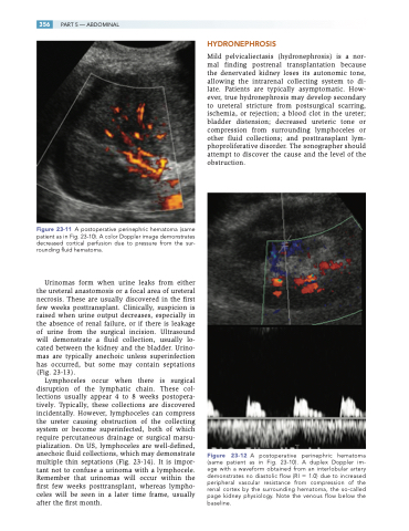
Figure 23-12 A postoperative perinephric hematoma (same patient as in Fig. 23-10). A duplex Doppler im- age with a waveform obtained from an interlobular artery demonstrates no diastolic flow (RI 1.0) due to increased peripheral vascular resistance from compression of the renal cortex by the surrounding hematoma, the so-called page kidney physiology. Note the venous flow below the baseline.
