
Page 375 - Libro 2
P. 375
23 — Evaluation of Kidney and Liver Transplants
355
Patients with graft dysfunction most commonly pres- ent with nonspecific signs and symptoms such as renal failure, pain, or evidence of infection. The goal of the US examination is to differentiate between causes of graft failure that are best managed medi- cally, such as acute tubular necrosis, pyelonephri- tis, drug toxicity, or rejection, from etiologies that require intervention such as hydronephrosis, symp- tomatic fluid collections, and vascular thromboses or stenosis. Unfortunately, many of the US findings in such patients, such as increase in renal length, loss of or increase in corticomedullary differentia- tion, striation of the uroepithelium, and increased RI are also nonspecific findings of graft dysfunction and diagnosis may ultimately require US-guided re- nal biopsy. However, Doppler criteria are highly spe- cific for most vascular complications following renal transplantation.
TRANSPLANT REJECTION
Rejection is one of the most common causes of graft loss and is the result of an attack by the im- mune system on the transplanted organ just as the immune system would combat any foreign object or virus. There are three types of renal transplant rejection: hyperacute, which occurs immediately postop due to the presence of preformed antibod- ies to the allograft; acute, which usually begins approximately 2 weeks posttransplantation, with most cases occurring in the first 3 months; and chronic. Fortunately, adjusting the immunosup- pression protocol can effectively treat most epi- sodes of rejection.
Rejection is suspected when one or more of the following clinical signs are detected: sudden cessa- tion of urine output called anuria, decreased urine output called oliguria, increase serum creatinine, protein or lymphocytes in the urine, hypertension, or swelling or tenderness of the graft. One of the earliest signs of rejection is oliguria, with an associated rise in serum creatinine and blood urea nitrogen (BUN). Serum creatinine and BUN determine how well the kidney is functioning because these waste products are normally removed from the blood by the kidneys. However, a rise in creatinine is a nonspecific finding and may indicate a variety of underlying renal pa- thologies. A biopsy should be performed in patients with a high level of creatinine that persists or contin- ues to increase.
ACUTE TUBULAR NECROSIS
Another common cause of graft dysfunction is ATN. ATN is caused by ischemia and is more common in DD transplants than in LRD. Risk factors for the de- velopment of ATN include prolonged ischemic time,
hypotension or blood loss during surgery, prolonged ICU time or severe illness of the donor, and harvest from a non-heart–beating donor. ATN occurs in the early postoperative period usually beginning day 2 or 3 and may be a cause for delayed function of the renal transplant. The patient may require dialysis until the kidney starts to function properly.7,8 Some investigators have used diminished diastolic flow in the segmental arteries as an indicator of ATN, whereas most use biopsy as the definitive diagnosis for ATN.9,10
FLUID COLLECTIONS
The most common perinephric fluid collections found in postrenal transplantation are hematomas, urinomas, and lymphoceles. The size and location of the collection should be documented on each ultra- sound examination.
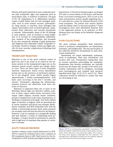
Hematomas are found immediately postopera- tively or postbiopsy. Their size, echotexture, and location will vary. Postoperative hematomas may be located anywhere surrounding the transplant. Hematomas that develop postbiopsy are typically found near the biopsy site, usually at the lower pole. Acutely, hematomas will be echogenic, becoming more heterogeneous and complex with anechoic liquefied areas (Figs. 23-10, 23-11, and 23-12). These collections should be followed to ensure that they are decreasing in size.
Figure 23-10 A postoperative perinephric hematoma. The grayscale sagittal image demonstrates a heterogeneous hypo- echoic fluid collection surrounding the kidney. The cortex of the lower pole appears compressed by this collection. The echogenicity or perinephric hematomas are variable, depend- ing on the time since the hemorrhage occurred.
