
Page 379 - Libro 2
P. 379
23 — Evaluation of Kidney and Liver Transplants
359
transplantation than following DD renal transplanta- tion. RAS may also occur secondary to twisting or kink- ing of the main renal artery. Excessive length of the renovascular pedicle predisposes a patient to vascular torsion.
To diagnose RAS on US examination, the entire length of the main renal artery and the anastomotic site must be carefully evaluated. Color Doppler should be optimized for assessing relatively high arterial ve- locities. The sonographer should use color Doppler to look for areas of aliasing with possible narrowing along the course of the main renal artery as well as for sharp bends or kinking of the artery. Any area of narrowing or aliasing should be sampled with spectral Doppler with an angle 60° and a sample volume just large enough to encompass the width of the re- nal artery. Doppler criteria for the diagnosis of RAS greater than 50% to 60% in a renal transplant include elevated peak systolic velocities greater than 200 to 250 cm/s, renal artery to external iliac artery ratio 2.0 to 3.0, plus poststenotic turbulence. In some patients, the distal arterial signal from the intraparen- chymal renal arteries may have a tardus–parvus wave- form pattern (Figs. 23-18 through 23-21).
Postbiopsy Vascular Complications
Patients who have had a renal biopsy may develop either an AVF or a pseudoaneurysm (PSA). An AVF is an abnormal connection between an artery and a vein.
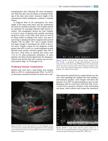
Figure 23-18 A renal artery stenosis. A color Doppler image of a renal artery anastomosis demonstrates focal color aliasing and a soft tissue color bruit (arrow). MRA, main renal artery; ILA, external iliac artery.
Figure 23-19 A renal artery stenosis (same patient as in Fig. 23-18). A grayscale image demonstrating narrowing (arrow) of the main renal artery (MRA) above the anastomosis, which was due to kinking of the vessel. Excessive length of the donor main renal artery predisposes to kinking of the vascular pedicle. ILA, external iliac artery.
This causes the arterial blood to empty directly into the vein, thus bypassing the capillary bed and creating a low-resistance gradient. Color Doppler will detect the presence of an AVF by demonstrating an area of color aliasing as well as a soft tissue color bruit (Fig. 23-22). A color bruit is caused by vibration of the surrounding soft tissue, which reflects back toward the transducer
Figure 23-20 A renal artery stenosis (same patient as in Fig. 23-18). A pulsed Doppler tracing obtained at the area of narrowing demonstrating increased PSV 440 cm/s.
