
Page 384 - Libro 2
P. 384
364
PART 5 — ABDOMINAL
Figure 23-29 A grayscale ultrasound of a piggyback IVC anas- tomosis. IVC-R, recipient IVC; HVC-R, recipient hepatic vein confluence; IVC-D, donor IVC.
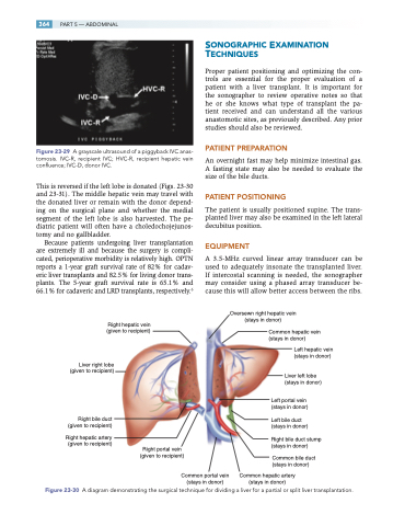
This is reversed if the left lobe is donated (Figs. 23-30 and 23-31). The middle hepatic vein may travel with the donated liver or remain with the donor depend- ing on the surgical plane and whether the medial segment of the left lobe is also harvested. The pe- diatric patient will often have a choledochojejunos- tomy and no gallbladder.
Because patients undergoing liver transplantation are extremely ill and because the surgery is compli- cated, perioperative morbidity is relatively high. OPTN reports a 1-year graft survival rate of 82% for cadav- eric liver transplants and 82.5% for living donor trans- plants. The 5-year graft survival rate is 65.1% and 66.1% for cadaveric and LRD transplants, respectively.6
Right hepatic vein (given to recipient)
SONOGRAPHIC EXAMINATION TECHNIQUES
Proper patient positioning and optimizing the con- trols are essential for the proper evaluation of a patient with a liver transplant. It is important for the sonographer to review operative notes so that he or she knows what type of transplant the pa- tient received and can understand all the various anastomotic sites, as previously described. Any prior studies should also be reviewed.
PATIENT PREPARATION
An overnight fast may help minimize intestinal gas. A fasting state may also be needed to evaluate the size of the bile ducts.
PATIENT POSITIONING
The patient is usually positioned supine. The trans- planted liver may also be examined in the left lateral decubitus position.
EQUIPMENT
A 3.5-MHz curved linear array transducer can be used to adequately insonate the transplanted liver. If intercostal scanning is needed, the sonographer may consider using a phased array transducer be- cause this will allow better access between the ribs.
Oversewn right hepatic vein (stays in donor)
Common hepatic vein (stays in donor)
Left hepatic vein (stays in donor)
Liver left lobe (stays in donor)
Left portal vein (stays in donor)
Left bile duct (stays in donor)
Right bile duct stump (stays in donor)
Common bile duct (stays in donor)
Common hepatic artery (stays in donor)
Liver right lobe (given to recipient)
Right bile duct (given to recipient)
Right hepatic artery (given to recipient)
Right portal vein (given to recipient)
Common portal vein (stays in donor)
Figure 23-30 A diagram demonstrating the surgical technique for dividing a liver for a partial or split liver transplantation.
