
Page 386 - Libro 2
P. 386
366
PART 5 — ABDOMINAL
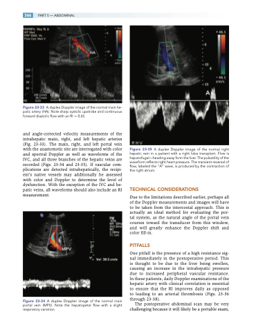
Figure 23-33 A duplex Doppler image of the normal main he- patic artery (HA). Note sharp systolic upstroke and continuous forward diastolic flow with an RI 0.65.
and angle-corrected velocity measurements of the intrahepatic main, right, and left hepatic arteries (Fig. 23-33). The main, right, and left portal vein with the anastomotic site are interrogated with color and spectral Doppler as well as waveforms of the IVC, and all three branches of the hepatic veins are recorded (Figs. 23-34 and 23-35). If vascular com- plications are detected intrahepatically, the recipi- ent’s native vessels may additionally be assessed with color and Doppler to determine the level of dysfunction. With the exception of the IVC and he- patic veins, all waveforms should also include an RI measurement.
Figure 23-34 A duplex Doppler image of the normal main portal vein (MPV). Note the hepatopetal flow with a slight respiratory variation.
Figure 23-35 A duplex Doppler image of the normal right hepatic vein in a patient with a right lobe transplant. Flow is hepatofugal—heading away form the liver. The pulsatility of the waveform reflects right heart pressure. The transient reversal of flow, labeled the “A” wave, is produced by the contraction of the right atrium.
TECHNICAL CONSIDERATIONS
Due to the limitations described earlier, perhaps all of the Doppler measurements and images will have to be taken from the intercostal approach. This is actually an ideal method for evaluating the por- tal system, as the natural angle of the portal vein courses toward the transducer from this window, and will greatly enhance the Doppler shift and color fill-in.
PITFALLS
One pitfall is the presence of a high resistance sig- nal immediately in the postoperative period. This is thought to be due to the liver being swollen, causing an increase in the intrahepatic pressure due to increased peripheral vascular resistance. In these patients, daily Doppler examinations of the hepatic artery with clinical correlation is essential to ensure that the RI improves daily as opposed to leading to an arterial thrombosis (Figs. 23-36 through 23-38).
The postoperative abdominal scan may be very challenging because it will likely be a portable exam,
