
Page 388 - Libro 2
P. 388
368 PART 5 — ABDOMINAL TABLE 23-4
Normal Doppler Findings Post–Liver Transplantation11,12
Vessel Direction/Color Normal Doppler Values
Main portal vein Right portal vein Left portal vein Main hepatic artery Right hepatic artery Left hepatic artery IVC
Hepatic veins
Hepatopetal/above baseline/red Hepatofugal/below baseline/blue Hepatopetal/above baseline/red Hepatopetal/above baseline/red Hepatofugal/below baseline/blue Hepatopetal/above baseline/red Can be bidirectional/pulsatile
hepatofugal/below baseline/blue (can also be slightly pulsatile due to proximity of heart)
125 cm/s stenosis, respiratory variations Forward, continuous flow
Forward continuous flow
RI 0.50, AT 80 ms, velocity 200 cm/s Same
Same
Velocity not measured
Velocity not measured
such as free abdominal fluid, periadrenal collections, and hematomas, are also documented. Indirect so- nographic signs of vascular complications may be seen in the liver parenchyma. These are frequently infarcts due to vascular insufficiency.
COMMON POSTOPERATIVE VASCULAR COMPLICATIONS
Table 23-6 lists the common vascular complications following liver transplantation. Using duplex tech- niques, one may see color filling defects when a
TABLE 23-5
Common Nonvascular Postop Liver Transplantation Complications
Bile duct obstruction Anastomotic bile duct obstruction Anastomotic stenosis/stricture Stone formation
Bile leak/biloma
Biliary necrosis
Cholangitis
Postoperative bleeding Hematoma
Abscess
Infection
Recurrent hepatitis
Portal hypertension
Splenic infarct
Recurrent malignancy Lymphoproliferative disorder
thrombus is present, color aliasing and spectral broadening with stenosis, or a complete or partial absence of flow with thrombi. The presence of vas- cular findings on a US examination may precipitate further imaging studies such as angiography, com- puterized tomography, and subsequent interven- tional procedures. Hepatic artery complications are a cause for immediate surgical intervention because the hepatic artery is the sole blood supply to the bile ducts after transplantation and a lack of blood flow will lead to biliary necrosis and loss of the transplant.
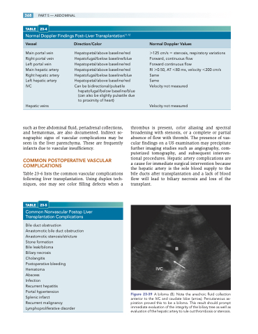
Figure 23-39 A biloma (B). Note the anechoic fluid collection anterior to the IVC and caudate lobe (arrow). Percutaneous as- piration proved this to be a biloma. This result should prompt immediate evaluation of the integrity of the biliary tree as well as evaluation of the hepatic artery to rule out thrombosis or stenosis.
