
Page 390 - Libro 2
P. 390
370
PART 5 — ABDOMINAL
Figure 23-42 A hepatic artery stenosis. A grayscale image of the porta hepatis demonstrating narrowing of the hepatic artery (arrow). This was believed to be due to poor surgical technique.
fever, biliary colic, or signs of hemorrhage. The Dop- pler waveform is often disorganized, showing a typi- cal mix of arterial and venous signal. There is a high risk of hemorrhage and resulting organ failure, so an interventional procedure must be performed to correct the PSA.
Figure 23-43 A hepatic artery stenosis (same patient as in Fig. 23-42). A spectral Doppler tracing demonstrates increased PSV 465 cm/s and turbulent flow. Notice the use of the color aliasing to guide placement of the Doppler cursor.
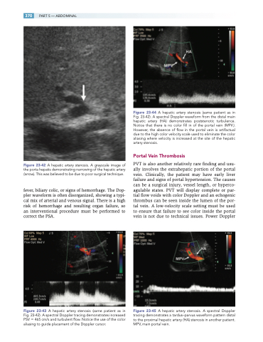
Figure 23-44 A hepatic artery stenosis (same patient as in Fig. 23-42). A spectral Doppler waveform from the distal main hepatic artery (HA) demonstrates poststenotic turbulence. Notice that there is no color fill in of the portal vein (MPV ). However, the absence of flow in the portal vein is artifactual due to the high color velocity scale used to eliminate the color aliasing where velocity is increased at the site of the hepatic artery stenosis.
Portal Vein Thrombosis
PVT is also another relatively rare finding and usu- ally involves the extrahepatic portion of the portal vein. Clinically, the patient may have early liver failure and signs of portal hypertension. The causes can be a surgical injury, vessel length, or hyperco- agulable states. PVT will display complete or par- tial flow voids with color Doppler and an echogenic thrombus can be seen inside the lumen of the por- tal vein. A low-velocity scale setting must be used to ensure that failure to see color inside the portal vein is not due to technical issues. Power Doppler
Figure 23-45 A hepatic artery stenosis. A spectral Doppler tracing demonstrates a tardus–parvus waveform pattern distal to the proximal hepatic artery (HA) stenosis in another patient. MPV, main portal vein.
