
Page 391 - Libro 2
P. 391
should also be used to verify absence of flow. An intervention is required to save the transplant and the bowel.
Portal Vein Stenosis
PVS is likely seen at the anastomotic sites and is often related to surgical injury. The patient may present with signs of worsening hepatic function that correlates with the degree of stenosis. On color and Doppler, an area of narrowing is easily identified in this large vessel. Also, a peak velocity at the area of greatest stenosis will be 125 cm/s or will have an anastomotic-to-preanastomotic ve- locity ratio of 3:1. An angioplasty or stent place- ment will be required.
23 — Evaluation of Kidney and Liver Transplants 371 IVC Thrombosis/Stenosis
Although rare, thrombosis or stenosis of the IVC is also a finding that is particularly relevant to the surgi- cal technique used to connect the recipient and donor IVCs. This finding is also associated with mechanical compression from fluid collections, hypercoagulability, vessel length, and retransplantation. The patient will often present with hepatic failure. It is helpful to know the technique used so that all portions of the IVC can be sampled. A thrombus can be seen inside the lumen, as well as visible signs of narrowing and velocity changes.
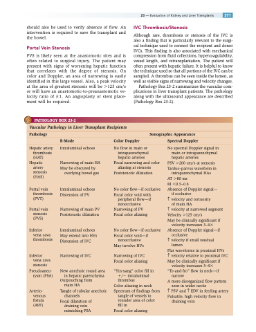
Pathology Box 23-2 summarizes the vascular com- plications in liver transplant patients. The pathology along with the ultrasound appearance are described (Pathology Box 23-2).
PATHOLOGY BOX 23-2
Vascular Pathology in Liver Transplant Recipients
Pathology Sonographic Appearance
B-Mode Color Doppler Spectral Doppler
Hepatic artery thrombosis (HAT)
Hepatic artery
stenosis (HAS)
Portal vein thrombosis (PVT)
Portal vein stenosis
(PVS)
Inferior
vena cava thrombosis
Inferior vena cava stenosis
Pseudoaneu- rysm (PSA)
Arterio- venous
fistula (AVF)
Intraluminal echoes
Narrowing of main HA May be obscured by
overlying bowel gas
Intraluminal echoes Distension of PV
Narrowing of main PV Poststenotic dilatation
Intraluminal echoes May extend into HVs Distension of IVC
Narrowing of IVC
New anechoic round area in hepatic parenchyma
Outpouching from main HA
Tangle of tubular anechoic channels
Focal dilatation of draining vein mimicking PSA
No flow in main or intraparenchymal hepatic arteries
Focal narrowing and color aliasing at stenosis
Poststenotic dilatation
No color flow—if occlusive Focal color void with
peripheral flow—if
nonocclusive Narrowing of PV Focal color aliasing
No color flow—if occlusive Focal color void—if
nonocclusive May involve HVs
Narrowing of IVC Focal color aliasing
“Yin-yang” color fill in / intraluminal thrombus
Color aliasing in neck Spectrum of findings from
tangle of vessels to rounder area of color fill in
Focal color aliasing
No spectral Doppler signal in main or intraparenchymal hepatic arteries
PSV 200 cm/s at stenosis Tardus–parvus waveform in intraparenchymal HAs
AT 80 ms
RI 0.5–0.6
Absence of Doppler signal—
if occlusive
↑ velocity and tortuosity
of main HA
↑ velocity at narrowed segment Velocity 125 cm/s
May be clinically significant if
velocity increases 3–4 Absence of Doppler signal—if
occlusive
↑ velocity if small residual
lumen
Flat waveforms in proximal HVs
↑ velocity relative to proximal IVC May be clinically significant if
velocity increases 3–4 “To-and-fro” flow in neck—if
narrow
A more disorganized flow pattern
seen in wider necks
↑ PSV and ↑ EDV in feeding artery Pulsatile, high velocity flow in
draining vein
