
Page 389 - Libro 2
P. 389
23 — Evaluation of Kidney and Liver Transplants 369
TABLE 23-6
Common Vascular Postop Liver Transplantation Complications
Hepatic artery thrombosis Hepatic artery stenosis Pseudoaneurysm
Portal vein thrombosis Portal vein stenosis
IVC thrombosis
IVC stenosis
Hepatic vein thrombosis
Hepatic vein stenosis
Biliary ischemia due to hepatic artery stenosis
Hepatic Artery Thrombosis
HAT is the most common vascular complication of the OLT. It occurs in 2% to 12% of liver transplants.11 The risk factors of developing HAT are rejection, pro- longed transport time of the organ, and use of an end-to-end surgical technique of the hepatic artery. Usually, the US examination will reveal absent or
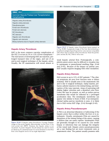
Figure 23-40 A hepatic artery thrombosis. A power Doppler image at the porta hepatis demonstrates normal flow in the main portal vein (MPV). However, the hepatic artery is not visualized. A power Doppler should always be used to show an absence of a vessel because it is more sensitive to slow flow than color Doppler and is not as angle dependent.
Figure 23-41 A hepatic artery thrombosis (same patient as in Fig. 23-40). A grayscale image of the liver demonstrating a peripheral wedge-shaped hypoechoic area (arrow) compatible with infarct due to hepatic artery thrombosis. Note the perihe- patic ascites (A). IVC, inferior vena cava.
weak hepatic arterial flow. Postsurgically, a com- pletely patent artery may be difficult to visualize due to vasospasm or parenchymal swelling (Figs. 23-40 and 23-41). Because of the impact on patient care, HAT usually requires imaging with other modalities.
Hepatic Artery Stenosis
HAS occurs in up to 11% of OLT patients.12 The clini- cal symptoms are poor liver function tests or biliary ischemia. HAS is usually seen at the anastomotic site and is due to surgical technique, clamp injuries, per- fusion catheter injuries to the intimal lining, and inter- ruption of the vasa vasorum. Areas of narrowing will display higher velocities and a disturbed color flow. Lower RIs may be present, as well as tardus–parvus waveforms that would be reflected by a prolonged acceleration time. The intrahepatic waveforms can appear very similar to those of HAS. If an interparen- chymal tardus–parvus waveform is seen, it is likely due to HAS versus HAT (Figs. 23-42 through 23-45).
Hepatic Artery Pseuodaneurysm
A PSA is an abnormal dilatation or ballooning of the hepatic artery that is either within the liver or is ex- trahepatic. Usually, extrahepatic PSAs are caused by disruption of the intimal lining of the artery, causing a dilation that can easily rupture. Intrahepatic PSAs are generally thought to be caused by core needle biopsy or infections that can damage the integrity of the vessel wall. Hepatic artery pseudoaneurysms are a rare finding but are more commonly seen at anastomotic sites. The patient usually presents with
