
Page 387 - Libro 2
P. 387
23 — Evaluation of Kidney and Liver Transplants
367
Figure 23-36 A duplex Doppler image of the main he- patic artery (HA) immediately posttransplantation demon- strates complete absence of diastolic flow and an RI 1.0. This high resistance waveform pattern is likely secondary to increased peripheral vascular resistance, which is second- ary to edema of the hepatic parenchyma. Within 48 hours posttransplantation, such a waveform pattern is not indica- tive of impending hepatic artery thrombosis. MPV, main portal vein.
and the scanning environment may be suboptimal. Also, the patient will likely be using an automated breathing apparatus, have multiple lines, and will likely have a completely bandaged abdomen. Of- ten, vacuum-assisted closure devices with foam centers are used for very large body cavity open- ings. An intercostal scanning technique may be the only method for sonographically evaluating the
Figure 23-37 A spectral Doppler tracing of the main hepatic artery (HA) 2 days later from the same patient as in Figure 23-36, demonstrating an increase in the amount of diastolic flow. The RI has dropped to 0.85. MPV, main portal vein.
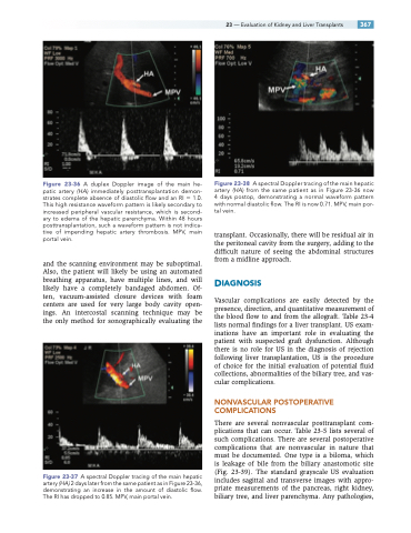
Figure23-38 AspectralDopplertracingofthemainhepatic artery (HA) from the same patient as in Figure 23-36 now 4 days postop, demonstrating a normal waveform pattern with normal diastolic flow. The RI is now 0.71. MPV, main por- tal vein.
transplant. Occasionally, there will be residual air in the peritoneal cavity from the surgery, adding to the difficult nature of seeing the abdominal structures from a midline approach.
DIAGNOSIS
Vascular complications are easily detected by the presence, direction, and quantitative measurement of the blood flow to and from the allograft. Table 23-4 lists normal findings for a liver transplant. US exam- inations have an important role in evaluating the patient with suspected graft dysfunction. Although there is no role for US in the diagnosis of rejection following liver transplantation, US is the procedure of choice for the initial evaluation of potential fluid collections, abnormalities of the biliary tree, and vas- cular complications.
NONVASCULAR POSTOPERATIVE COMPLICATIONS
There are several nonvascular posttransplant com- plications that can occur. Table 23-5 lists several of such complications. There are several postoperative complications that are nonvascular in nature that must be documented. One type is a biloma, which is leakage of bile from the biliary anastomotic site (Fig. 23-39). The standard grayscale US evaluation includes sagittal and transverse images with appro- priate measurements of the pancreas, right kidney, biliary tree, and liver parenchyma. Any pathologies,
