
Page 406 - Libro 2
P. 406
386
PART 6 — MISCELLANEOUS
catheter resides in a central vein, most often the superior vena cava. There are many different types of central VADs, each with varying characteristics. The goal for device selection is to match the right device to the right patient, with consideration given to therapy duration, number and type of infusions and patient lifestyle and activity issues. A thorough assessment prior to device placement is critical in selecting the right device and placing it in the right location for the right therapy.
Today, it is virtually the standard of care to use ultrasound guidance to assess potential target sites for VAD placement and to guide the initial venous puncture as the first step in device placement. The Agency for Healthcare Research and Quality has recommended the use of ultrasound as one of their 11 practices to improve patient care in their land- mark 2001 publication, “Making Health Care Safer: A Critical Analysis of Patient Safety Practices.”1,2 This chapter will focus on the role ultrasound im- aging plays in the placement of central vascular ac- cess devices.
ANATOMY
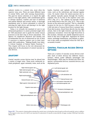
Central vascular access devices may be placed into a variety of target veins. These most commonly in- clude peripheral; upper extremity veins, such as the
Right internal jugular vein Right external jugular vein Right subclavian vein
Right brachiocephalic vein
Internal thoracic vein Right superior intercostal vein
Superior vena cava
Azygos vein Right pericardiophrenic vein
Inferior vena cava
basilic, brachial, and cephalic veins; and central veins, such as the subclavian and internal jugular veins. No matter where the initial puncture site is, the catheter typically passes through the brachio- cephalic vein on its way to the superior vena cava (SVC) (Fig. 25-1). The catheter tip typically resides in the distal third of the SVC (Fig. 25-2). This is also known as the atriocaval junction. This is a desirable location for the tip of a VAD because a flow rate of 2,000 mL/min of venous blood flow is present here. From this location, blood flows directly into the right atrium, then to the right ventricle, and then into the pulmonary circulation. Given the high blood flow in this location, infusates are rapidly dispersed and di- luted in this area of high flow. Aspiration for blood return, exchange transfusions, and dialysis or apher- esis is also easily accomplished when the VAD tip is accurately located.
CENTRAL VASCULAR ACCESS DEVICE OPTIONS
There are a variety of vascular access devices avail- able. Each of these devices has different charac- teristics, which confer different advantages and disadvantages. VADs may be divided into three cat- egories: nontunneled devices, tunneled devices, and implanted ports.
Left internal jugular vein Left external jugular vein
Left subclavian vein
Internal thoracic vein
Left superior intercostal vein Left brachiocephalic vein
Accessory hemiazygos vein Left pericardiophrenic vein
Hemiazygos vein
Figure25-1 Thisanatomicdrawingdemonstratesthecentralveins,includingtheinternaljugularandbrachiocephalicveinsinaddition to the superior vena cava, all of which are important for placement of vascular access devices. (Image courtesy of Michael Ciarmiello, Albany, NY.)
