
Page 407 - Libro 2
P. 407
25 — The Role of Ultrasound in Central Vascular Access Device Placement
387
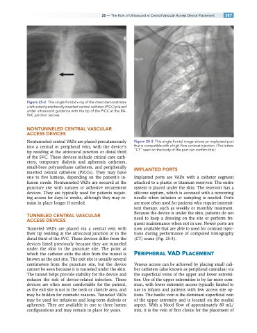
Figure 25-2 This single frontal x-ray of the chest demonstrates a left-sided peripherally inserted central catheter (PICC) placed under ultrasound guidance with the tip of the PICC at the RA- SVC junction (arrow).
NONTUNNELED CENTRAL VASCULAR ACCESS DEVICES
Nontunneled central VADs are placed percutaneously into a central or peripheral vein, with the device’s tip residing at the atriocaval junction or distal third of the SVC. These devices include critical care cath- eters, temporary dialysis and apheresis catheters, small-bore polyurethane catheters, and peripherally inserted central catheters (PICCs). They may have one to five lumens, depending on the patient’s in- fusion needs. Nontunneled VADs are secured at the puncture site with sutures or adhesive securement devices. They are typically used for patients requir- ing access for days to weeks, although they may re- main in place longer if needed.
TUNNELED CENTRAL VASCULAR ACCESS DEVICES
Tunneled VADs are placed via a central vein with their tip residing at the atriocaval junction or in the distal third of the SVC. These devices differ from the devices listed previously because they are tunneled under the skin to the puncture site. The point at which the catheter exits the skin from the tunnel is known as the exit site. The exit site is usually several centimeters from the puncture site, but the device cannot be seen because it is tunneled under the skin. The tunnel helps provide stability for the device and reduces the risk of device-related infection. These devices are often more comfortable for the patient, as the exit site is not in the neck or clavicle area, and may be hidden for cosmetic reasons. Tunneled VADs may be used for infusions and long-term dialysis or apheresis. They are available in one to three lumen configurations and may remain in place for years.
Figure 25-3 This single frontal image shows an implanted port that is compatible with a high-flow contrast injection. (The letters “CT” seen on the body of the port can confirm this.)
IMPLANTED PORTS
Implanted ports are VADs with a catheter segment attached to a plastic or titanium reservoir. The entire system is placed under the skin. The reservoir has a silicone septum, which is accessed with a noncoring needle when infusion or sampling is needed. Ports are most often used for patients who require intermit- tent therapy, such as weekly or monthly treatment. Because the device is under the skin, patients do not need to keep a dressing on the site or perform fre- quent maintenance when not in use. Newer ports are now available that are able to used for contrast injec- tions during performance of computed tomography (CT) scans (Fig. 25-3).
PERIPHERAL VAD PLACEMENT
Venous access can be achieved by placing small cali- ber catheters (also known as peripheral cannulas) via the superficial veins of the upper and lower extremi- ties. Use of the upper extremities is by far more com- mon, with lower extremity access typically limited to use in infants and patients with few access site op- tions. The basilic vein is the dominant superficial vein of the upper extremity and is located on the medial aspect. With a blood flow of approximately 80 mL/ min, it is the vein of first choice for the placement of
