
Page 408 - Libro 2
P. 408
388
PART 6 — MISCELLANEOUS
PICCs. The basilic vein drains directly into the axil- lary vein, which becomes the subclavian vein as it enters the chest. The brachial veins are also located medially in the upper extremity and are paired veins in proximity to the brachial artery. Typically smaller than the basilic vein, they are a good choice for ini- tial access as well. Their location adjacent to the brachial artery imparts a higher risk of inadvertent arterial puncture than other upper extremity veins. The cephalic vein is the smallest of the named upper extremity veins with a blood flow of approximately 30 mL/min and is thus the least preferred choice for VAD placement. It is positioned more laterally on the arm, making it easy to access. It joins the subclavian vein just past the shoulder.
Lower extremity veins, such as the saphenous vein or veins of the feet, may be used when there are no suitable upper extremity veins for access. This is more common in neonates and children. Although most pe- ripheral veins are palpable and visible, ultrasound may be useful to place peripheral cannulas in challenging patients and in the brachial or saphenous veins. Use of peripheral cannulas should be limited to very short- term therapies (less than 1 week). Peripheral cannulas are changed every 72 hours if possible to reduce the risk of vein damage and infiltration of infusates. Only infusates that are nonirritating and lack vesicant prop- erties should be administered via peripheral cannulas.
CENTRAL VAD PLACEMENT
The most common sites for central venous access are the internal jugular veins (IJVs) and the subcla- vian veins (SCVs). The IJVs are relatively superficial, facilitating assessment and cannulation. The right IJV approach is preferred over the left because it has a straighter course to the heart, making the procedure technically easier. In proximity to the internal jugu- lar veins are the smaller external and anterior jugular veins, which drain blood from the face and neck. The external jugular veins are superficial and often tortu- ous. They join the IJV at the confluence of the subcla- vian and brachiocephalic veins. Because of their size, they are not preferred for central venous access and are more often used in the setting of IJV occlusion.
SONOGRAPHIC EXAMINATION TECHNIQUES
Ultrasound allows for the anatomical assessment of the IJV prior to the procedure, as well as dynamic guidance during vein puncture. The IJV can be can- nulated blindly using an anatomical landmark ap- proach, which was and still is a common method for
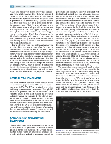
performing this procedure. However, compared with the landmark method, real-time ultrasound guidance has been shown to be both a quicker and safer way to accomplish this goal. Two-dimensional ultrasound guidance can reduce the failure of catheter placement and complication rates related to insertion by 86% and 57%, respectively.3 When using ultrasound, it is important to assess the depth of the vessel from the skin, the vessel patency, vessel diameter, variations in diameter with respiration, and the relationship of the vein to the common carotid artery (CCA). It is impor- tant to identify the CCA prior to attempted cannulation of the IJV. Typically, the IJV is located anterior and lat- eral to the CCA (Fig. 25-4). However, there are several variations of this relationship that can be encountered. In a prospective evaluation of 869 patients who had undergone real-time ultrasound-guided cannulation of the IJV, five anatomical arrangements of the IJV and CCA were found in 659 patients. In 328 cases (49.8%), the IJV was anterolateral to the CCA, whereas in an ad- ditional 146 cases (22.2%), it was lateral to the CCA. In 148 cases (22.5%), the vein lay directly anterior to the artery. In the remaining cases, the IJV was an- teromedial to the CCA in 30 cases (4.5%) and directly medial to the artery in seven cases (1.0%).4
The subclavian veins are commonly used to attain central venous access. The SCVs are located in the chest, adjacent to the subclavian arteries. They usually lie directly under the clavicle. Because of this location, they are more difficult to visualize with ultrasound than other target veins. SCVs are typically large and thus suitable for cannulation with larger bore central VADs, such as dialysis and apheresis catheters. They terminate in the brachiocephalic veins, at the conflu- ence with the internal jugular veins. Ultimately, the right and left brachiocephalic veins join to form the SVC. Neither the subclavian nor the upper extremity
Figure 25-4 This transverse image of the left side of the neck shows both the larger internal jugular vein anterior and slightly lateral to the smaller common carotid artery.
