
Page 409 - Libro 2
P. 409
25 — The Role of Ultrasound in Central Vascular Access Device Placement
389
veins should be used for cannulation in patients with chronic renal insufficiency or chronic kidney disease. Because use of these veins for VAD placement may be associated with thrombus and stenosis, avoidance of these veins preserves them for use as a permanent access for hemodialysis (arteriovenous fistula or graft formation) in the future.5
The common femoral veins (CFVs) are located in the groin, medial to the common femoral artery. The common femoral veins drain blood from the lower extremities into the external iliac veins. These ultimate- ly become the common iliac veins, which join to form the inferior vena cava. The CFVs are most often used for central venous access in emergent situations and in patients in whom other potential access veins are oc- cluded. VAD placement via the CFV is associated with a higher rate of mechanical and infectious complications, and therefore should be avoided unless necessary.
SCANNING TECHNIQUE
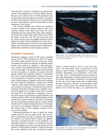
Ultrasound imaging is used to both assess potential access sites for VAD placement, as well as to guide the initial needle puncture into the access site. Initial assessment includes the evaluation of available patent vessels, their location in relation to other structures (e.g., arteries, other implanted devices), and the ability to access the target. The specific target vein is assessed for size and patency. A stenotic (or scarred) vein will appear smaller than expected. Patency is determined by a test of vessel compressibility, similar to that used in the assessment of a patient for deep venous throm- bosis. Gentle pressure is applied on the skin overlying the vein using the transducer. A patent vein should compress easily under pressure and should re-expand once pressure is released. A thrombosed vein will not compress using this technique and, in addition, its lu- men will appear more echogenic than that of a patent vein (Fig. 25-5). In comparison, compression of an ar- tery will result in less reduction of vessel diameter, and pulsation will be visible. Clearly differentiating venous vessels from arterial vessels is a key factor in reducing the risk of inadvertent arterial puncture. Vessel caliber should also be assessed using ultrasound. The target vein must be of adequate diameter to accommodate the selected VAD, so that complete occlusion of the vein can be avoided.
Ultrasound guidance is used to guide the initial needle puncture into the target vessel. Once the pa- tient is positioned, the skin is prepped with antisep- tic solution, and sterile barrier drapes are applied. The skin is anesthetized with a local anesthetic. Ad- ditionally, systemic sedation or anesthesia may be used depending on the type of device placed and the patient’s overall clinical condition. Sterile ultrasound gel is then applied to the skin overlying the target
Figure 25-5 This longitudinal image of the neck shows a non- compressible internal jugular vein and its relationship to the common carotid artery.
vessel. A sterile sheath is used to cover the trans- ducer. The ultrasound probe is positioned on the patient’s skin in either a longitudinal or transverse position, depending on the preference of the clini- cian (Fig. 25-6). A needle guide may be attached to the probe depending on clinician preference as well. The depth of the probe is adjusted to accommodate for the size of the target vessel and its depth below the skin. Typically, a small (21 g) hyperechoic needle is used to attain access. The needle should be visu- alized once it enters the skin, as it approaches the
Figure 25-6 This single image during VAD placement shows the longitudinal approach to venous access.
