
Page 410 - Libro 2
P. 410
390 PART 6 — MISCELLANEOUS
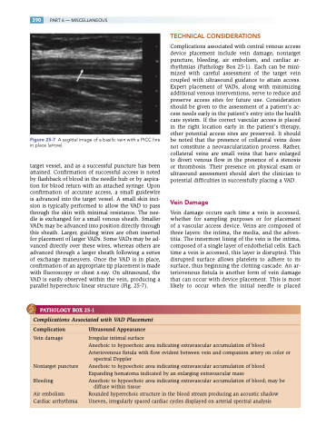
Figure 25-7 A sagittal image of a basilic vein with a PICC line in place (arrow).
target vessel, and as a successful puncture has been attained. Confirmation of successful access is noted by flashback of blood in the needle hub or by aspira- tion for blood return with an attached syringe. Upon confirmation of accurate access, a small guidewire is advanced into the target vessel. A small skin inci- sion is typically performed to allow the VAD to pass through the skin with minimal resistance. The nee- dle is exchanged for a small venous sheath. Smaller VADs may be advanced into position directly through this sheath. Larger, guiding wires are often inserted for placement of larger VADs. Some VADs may be ad- vanced directly over these wires, whereas others are advanced through a larger sheath following a series of exchange maneuvers. Once the VAD is in place, confirmation of an appropriate tip placement is made with fluoroscopy or chest x-ray. On ultrasound, the VAD is easily observed within the vein, producing a parallel hyperechoic linear structure (Fig. 25-7).
PATHOLOGY BOX 25-1
TECHNICAL CONSIDERATIONS
Complications associated with central venous access device placement include vein damage, nontarget puncture, bleeding, air embolism, and cardiac ar- rhythmias (Pathology Box 25-1). Each can be mini- mized with careful assessment of the target vein coupled with ultrasound guidance to attain access. Expert placement of VADs, along with minimizing additional venous interventions, serve to reduce and preserve access sites for future use. Consideration should be given to the assessment of a patient’s ac- cess needs early in the patient’s entry into the health care system. If the correct vascular access is placed in the right location early in the patient’s therapy, other potential access sites are preserved. It should be noted that the presence of collateral veins does not constitute a neovascularization process. Rather, collateral veins are small veins that have enlarged to divert venous flow in the presence of a stenosis or thrombosis. Their presence on physical exam or ultrasound assessment should alert the clinician to potential difficulties in successfully placing a VAD.
Vein Damage
Vein damage occurs each time a vein is accessed, whether for sampling purposes or for placement of a vascular access device. Veins are composed of three layers: the intima, the media, and the adven- titia. The innermost lining of the vein is the intima, composed of a single layer of endothelial cells. Each time a vein is accessed, this layer is disrupted. This disrupted surface allows platelets to adhere to its surface, thus beginning the clotting cascade. An ar- teriovenous fistula is another form of vein damage that can occur with device placement. This is most likely to occur when the initial needle is placed
Complications Associated with VAD Placement
Complication UltrasoundAppearance
Vein damage
Nontarget puncture Bleeding
Air embolism Cardiac arrhythmia
Irregular intimal surface
Anechoic to hypoechoic area indicating extravascular accumulation of blood Arteriovenous fistula with flow evident between vein and companion artery on color or
spectral Doppler
Anechoic to hypoechoic area indicating extravascular accumulation of blood Expanding hematoma indicated by an enlarging extravascular mass
Anechoic to hypoechoic area indicating extravascular accumulation of blood; may be
diffuse within tissue
Rounded hyperechoic structure in the blood stream producing an acoustic shadow Uneven, irregularly spaced cardiac cycles displayed on arterial spectral analysis
