
Page 414 - Libro 2
P. 414
394
PART 6 — MISCELLANEOUS
AV fistulae have 2-year primary patency rates of 40% to 69%. In comparison, 2-year primary patency rates for prosthetic grafts range from 18% to 30%. Secondary patency rates for fistulae range from 62% to 75% at 2 years. For prosthetic grafts, secondary patency rates are 40% to 60% in the same time frame. The trade-off for higher long-term AV fistula patency rates is a lower maturation rate and higher early thrombosis rate. More often, maturation failure is due to the use of small or suboptimal veins.2
PREOPERATIVE EVALUATION
SONOGRAPHIC EXAMINATION TECHNIQUE
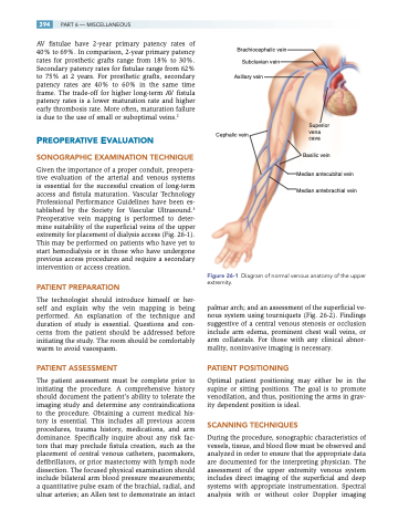
Given the importance of a proper conduit, preopera- tive evaluation of the arterial and venous systems is essential for the successful creation of long-term access and fistula maturation. Vascular Technology Professional Performance Guidelines have been es- tablished by the Society for Vascular Ultrasound.3 Preoperative vein mapping is performed to deter- mine suitability of the superficial veins of the upper extremity for placement of dialysis access (Fig. 26-1). This may be performed on patients who have yet to start hemodialysis or in those who have undergone previous access procedures and require a secondary intervention or access creation.
PATIENT PREPARATION
The technologist should introduce himself or her- self and explain why the vein mapping is being performed. An explanation of the technique and duration of study is essential. Questions and con- cerns from the patient should be addressed before initiating the study. The room should be comfortably warm to avoid vasospasm.
PATIENT ASSESSMENT
The patient assessment must be complete prior to initiating the procedure. A comprehensive history should document the patient’s ability to tolerate the imaging study and determine any contraindications to the procedure. Obtaining a current medical his- tory is essential. This includes all previous access procedures, trauma history, medications, and arm dominance. Specifically inquire about any risk fac- tors that may preclude fistula creation, such as the placement of central venous catheters, pacemakers, defibrillators, or prior mastectomy with lymph node dissection. The focused physical examination should include bilateral arm blood pressure measurements; a quantitative pulse exam of the brachial, radial, and ulnar arteries; an Allen test to demonstrate an intact
Brachiocephalic vein Subclavian vein
Axillary vein
Cephalic vein
Superior vena cava
Basilic vein
Median antecubital vein Median antebrachial vein
Figure 26-1 Diagram of normal venous anatomy of the upper extremity.
palmar arch; and an assessment of the superficial ve- nous system using tourniquets (Fig. 26-2). Findings suggestive of a central venous stenosis or occlusion include arm edema, prominent chest wall veins, or arm collaterals. For those with any clinical abnor- mality, noninvasive imaging is necessary.
PATIENT POSITIONING
Optimal patient positioning may either be in the supine or sitting positions. The goal is to promote venodilation, and thus, positioning the arms in grav- ity dependent position is ideal.
SCANNING TECHNIQUES
During the procedure, sonographic characteristics of vessels, tissue, and blood flow must be observed and analyzed in order to ensure that the appropriate data are documented for the interpreting physician. The assessment of the upper extremity venous system includes direct imaging of the superficial and deep systems with appropriate instrumentation. Spectral analysis with or without color Doppler imaging
