
Page 416 - Libro 2
P. 416
396
PART 6 — MISCELLANEOUS
Figure 26-5 Increased intraluminal echoes consistent with occluding thrombosis in a basilic vein.
needle puncture. Documentation of vessel character- istics should include patency, depth, wall thickness, calcification, and location of a thrombus or fibrosis.
For maximal vein dilation, a double tourniquet tech- nique can be used. One tourniquet should be placed on the forearm, which compresses the superficial ves- sels, and the other tourniquet should be placed in the axillary region to compress the deeper veins.
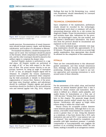
Spectral Doppler analysis is performed in a sag- ittal plane. All Doppler studies are performed at an angle of 60° or less with respect to the direc- tion of flow. The actual venous velocity is not im- portant; however, by using an angle of insonation of 60° or less, an adequate Doppler signal will be obtained. To complete the venous examination, spectral waveforms are performed while compress- ing the limb proximally or distally to demonstrate augmentation of venous flow. Patency of the proxi- mal deep veins (brachial, axillary, and subclavian) should be confirmed. Doppler signals and spectral waveforms should be checked in the mid-subclavian vein and internal jugular vein (Fig. 26-6). Atypical
Figure 26-6 Doppler imaging of the mid-subclavian vein showing respiratory phasicity.
findings that may be life threatening (e.g., central vein thrombosis) should immediately be conveyed to a health care provider.
TECHNICAL CONSIDERATIONS
Upon completion of the examination, preliminary results should be recorded by the technologist. A technologist work sheet or summary will aid the interpreting physician while he or she reviews the diagnostic images. Proper documentation is essential and includes exam date, indications for the proce- dure, the technologist’s name, the arm studied, and the patient’s identification information. Any devia- tion from the protocol should be documented and explained on the work sheet.
The routine unilateral upper extremity vein map- ping examination should take approximately 35 to 45 minutes. The goals are to provide an accurate high quality examination, thus appropriate time should be allowed for the completion of this examination.
PITFALLS
There are few contraindications to this ultrasound- guided assessment, and they include intravenous lines, dressings, open wounds, and limited patient positioning. Attempts should be made to use vari- ous approaches and scanning orientations to work around some of these obstacles.
DIAGNOSIS
For the assessment of the radial, ulnar, and brachial arteries, an arterial diameter greater than 2 mm is needed for fistula creation and maturation. Matu- ration is defined as a dilated, easily palpable, and usable fistula for hemodialysis at a flow rate of 350 mL/min.4 Volume flow rates will be discussed later in this chapter. The grayscale image should demonstrate smooth walled vessels that are free of disease. Calcification will appear as bright white echoes along or within the vessel walls.
Venous criteria for acceptable conduit diameter varies between physicians. Although there is no con- sensus, a favorable vessel diameter for creation of AV access is greater than 2.5 mm. Using this diameter as a minimum, it has been shown that a 92% early maturation rate and an 83% 1-year patency rate can be obtained.5 Vein walls should be completely com- pressible with light transducer pressure. A partially compressible or noncompressible vein suggests the presence of an occluding thrombus within the vein lumen, making it unusable as an autogenous conduit.
