
Page 418 - Libro 2
P. 418
398
PART 6 — MISCELLANEOUS
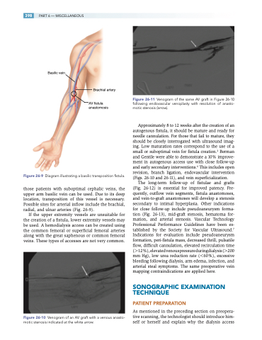
Basilic vein
Brachial artery
AV fistula anastomosis
Figure 26-11 Venogram of the same AV graft in Figure 26-10 following endovascular venoplasty with resolution of anasto- motic stenosis (arrow).
Approximately 8 to 12 weeks after the creation of an autogenous fistula, it should be mature and ready for needle cannulation. For those that fail to mature, they should be closely interrogated with ultrasound imag- ing. Low maturation rates correspond to the use of a small or suboptimal vein for fistula creation.2 Berman and Gentile were able to demonstrate a 10% improve- ment in autogenous access use with close follow-up and early secondary interventions.6 This includes open revision, branch ligation, endovascular intervention (Figs. 26-10 and 26-11), and vein superficialization.
The long-term follow-up of fistulae and grafts (Fig. 26-12) is essential for improved patency. Fre- quently, outflow vein segments, fistula anastomoses, and vein-to-graft anastomoses will develop a stenosis secondary to intimal hyperplasia. Other indications for close follow-up include pseudoaneurysm forma- tion (Fig. 26-13), mid-graft stenosis, hematoma for- mation, and arterial stenosis. Vascular Technology Professional Performance Guidelines have been es- tablished by the Society for Vascular Ultrasound.7 Indications for evaluation include pseudoaneurysm formation, peri-fistula mass, decreased thrill, pulsatile flow, difficult cannulation, elevated recirculation time (12%),elevatedvenouspressureduringdialysis(200 mm Hg), low urea reduction rate (60%), excessive bleeding following dialysis, arm edema, infection, and arterial steal symptoms. The same preoperative vein mapping contraindications are applied here.
SONOGRAPHIC EXAMINATION TECHNIQUE
PATIENT PREPARATION
As mentioned in the preceding section on preopera- tive scanning, the technologist should introduce him- self or herself and explain why the dialysis access
Figure 26-9 Diagram illustrating a basilic transposition fistula.
those patients with suboptimal cephalic veins, the upper arm basilic vein can be used. Due to its deep location, transposition of this vessel is necessary. Possible sites for arterial inflow include the brachial, radial, and ulnar arteries (Fig. 26-9).
If the upper extremity vessels are unsuitable for the creation of a fistula, lower extremity vessels may be used. A hemodialysis access can be created using the common femoral or superficial femoral arteries along with the great saphenous or common femoral veins. These types of accesses are not very common.
Figure 26-10 Venogram of an AV graft with a venous anasto- motic stenosis indicated at the white arrow.
