
Page 420 - Libro 2
P. 420
400
PART 6 — MISCELLANEOUS
for the interpreting physician. Spectral analysis with or without color Doppler imaging should be performed using a carrier frequency of at least 7 to 12 MHz. Sys- tem presets should be adjusted to the high flow set- tings, and decreasing color gain will minimize tissue bruit. Use B-mode to assess for various abnormalities such as perigraft masses, pseudoaneurysms, stenotic valves, and intimal flaps. Perigraft fluid collections and pseudoaneurysms can be differentiated using B-mode, color flow imaging, and Doppler imaging. The fistula diameter should be measured along its length. Scan- ning the fistula in a transverse view will locate side branches that can limit fistula maturation. Measure the branch vessel diameter and document the location.
Doppler evaluation is used to document patency of the fistula as well as to identify areas of stenosis. Dop- pler spectral analysis is performed in the sagittal plane. All Doppler samples must be performed at an angle of 60° or less with respect to the direction of flow. Doppler cursor alignment should be parallel to the vessel wall. When a stenosis is found, velocities should be mea- sured proximal, distal, and within the area of interest. Arterial inflow is first assessed with duplex imaging. Peak systolic velocities (PSVs) should be recorded in the native artery proximal to the anastomosis, at the anas- tomosis, throughout the body of the fistula, and along the venous outflow. Scanning throughout the entire fis- tula or graft, obtaining spectral signals, and paying close attention to the needle puncture sites is essential. The examination must include the venous outflow and, in some cases, may extend into the chest for assessing the central veins. Color imaging is helpful in this location.
Volume flow measurements are useful when eval- uating access function and are best accomplished with a wide-open sample gate. Volume flow calcula- tions should be made in the mid-graft or fistula at a site of normal flow. The volumetric flow (millimeters per minute) is calculated as follows:
Flow Time Average Velocity Area 60. The area is calculated based on:
(1/2 diameter)2.
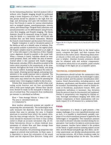
Most modern ultrasound systems are capable of measuring this automatically. The technologist man- ually measures the diameter, which the ultrasound system then uses to calculate the area. A Doppler spectrum is recorded from which the system calcu- lates the time average velocity. The time average ve- locity should be measured over three to four cardiac cycles to obtain an accurate calculation (Fig. 26-14). Some laboratories choose to obtain triplicate mea- surements of volume flow and then average these three values to improve accuracy.
In select situations, the presenting symptoms may suggest an arterial steal syndrome. In these cases, it is imperative to know the orientation of the sound beam and the expected flow direction. Using color
Figure 26-14
calculated.
A duplex ultrasound of a fistula with volume flow
flow, check for retrograde flow in the distal native artery, compress the graft, and then reassess flow direction in the native artery. Measuring distal arteri- al velocity or flow with and without fistula compres- sion is helpful. Absolute forearm pressures should also be obtained with and without compression. In situations when this is not possible, pulse volume recordings of the digits are an acceptable option.
TECHNICAL CONSIDERATIONS
Documentation should include the examination date, indications for the procedure, the technologist’s name, the arm studied, and the patient’s identification infor- mation. Vessel characteristics, including patency, wall thickness, calcification, and thrombus content, should be recorded. Documenting native vessel or fistula ste- nosis or thrombosis, anastomotic lesions, PSVs, and poststenotic turbulence is necessary. Any deviation from the protocol must be documented and explained. Similar to preoperative vein mapping for dialysis ac- cess, thorough surveillance imaging with direct and indirect components of these examinations will rou- tinely require 35 to 45 minutes for a unilateral limb.
Pitfalls
The examination of a fistula or graft presents a few unique challenges. It is a very superficial structure and, as such, it is possible to inadvertently partially com- press it if too much transducer pressure is applied by the technologist or sonographer. This transducer pressure can result in elevated velocities recorded during the ex- amination. Care needs to be taken to avoid excess trans- ducer pressure. Another factor related to the position of the fistula or graft involves scanning mature access sites. As both fistulae and grafts age, they dilate, can be- come aneurysmal, can develop pseudoaneurysms, and can become tortuous. All of these conditions make for an irregular scanning surface on the arm. The surface
