
Page 421 - Libro 2
P. 421
26 — Hemodialysis Access Grafts and Fistulae
401
Figure 26-15 Color flow imaging and spectral Doppler of a normally functioning fistula.
irregularities require regular, large amounts of gel to maintain proper skin contact. Larger pseudoaneurysms may require scanning from various approaches to fully document all the findings. Occasionally, patients pres- ent with contraindications to scanning including dress- ings or open wounds or possibly decreased mobility leading to poor patient positioning.
DIAGNOSIS
The grayscale image should reveal an access free of a thrombus or calcifications. A thrombus may appear hypoechoic or anechoic depending on its age. Calcifica- tions will appear as bright white reflectors with the ves- sel walls. In a fistula, the valves within the vein should not be evident and should appear to be adhered to the vessel wall. Any valve projecting into the lumen should be noted, as it can be a source for stenosis development.
A well-functioning fistula will have a PSV between 150 and 300 cm/s and end-diastolic velocities (EDVs) of 60 to 200 cm/s (Fig. 26-15).8 High-grade stenoses are present with focal velocity elevations of 100%, thus producing a velocity ratio (Vr) 2 as compared to the adjacent segment.9 A stenosis is also likely to be
Figure 26-16 Image of an anastomotic stenosis with elevated velocities.
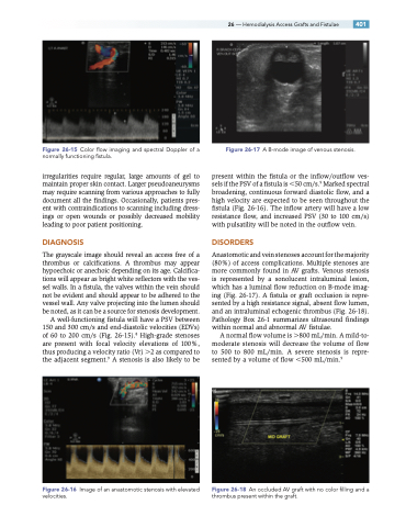
Figure 26-17 A B-mode image of venous stenosis.
present within the fistula or the inflow/outflow ves- sels if the PSV of a fistula is 50 cm/s.9 Marked spectral broadening, continuous forward diastolic flow, and a high velocity are expected to be seen throughout the fistula (Fig. 26-16). The inflow artery will have a low resistance flow, and increased PSV (30 to 100 cm/s) with pulsatility will be noted in the outflow vein.
DISORDERS
Anastomotic and vein stenoses account for the majority (80%) of access complications. Multiple stenoses are more commonly found in AV grafts. Venous stenosis is represented by a sonolucent intraluminal lesion, which has a luminal flow reduction on B-mode imag- ing (Fig. 26-17). A fistula or graft occlusion is repre- sented by a high resistance signal, absent flow lumen, and an intraluminal echogenic thrombus (Fig. 26-18). Pathology Box 26-1 summarizes ultrasound findings within normal and abnormal AV fistulae.
A normal flow volume is 800 mL/min. A mild-to- moderate stenosis will decrease the volume of flow to 500 to 800 mL/min. A severe stenosis is repre- sented by a volume of flow 500 mL/min.9
Figure 26-18 An occluded AV graft with no color filling and a thrombus present within the graft.
