
Page 419 - Libro 2
P. 419
26 — Hemodialysis Access Grafts and Fistulae
399
Graft
PATIENT ASSESSMENT
The patient assessment is similar to that used for the preoperative testing and should be completed prior to initiating the procedure. A comprehensive medical and surgical history should be documented including the patient’s ability to tolerate the imaging study. Any potential contraindications should be determined. The history should include all previous access procedures, trauma history, medications, and arm dominance. If available, reviewing access diagrams or operative notes will clarify the access type and location. Specifically, inquire about any risk factors that may preclude fistula maturation or use, such as central venous thrombosis or placement of central venous catheters, pacemak- ers, or defibrillators. The focused physical examination should include a quantitative exam of the fistula or graft. Assessment for the presence of a thrill, and the quality of the thrill throughout the entire access is im- portant. Visual inspection of the arm and access site is necessary to assess for edema, redness, presence of col- lateral veins, rotation of access sites, and focal dilations.
PATIENT POSITIONING
Optimal patient positioning should be supine, with the arm extended and relaxed. Depending on the exact placement of the dialysis access site, the arm position may need to be adjusted during the course of the examination.
SCANNING TECHNIQUES
The patient and arm are placed in a comfortable posi- tion. Use minimal pressure and abundant ultrasound gel to optimize imaging. Characteristics of vessels, tis- sue, and blood flow must be observed and analyzed in order to ensure that appropriate data is documented
Brachial artery
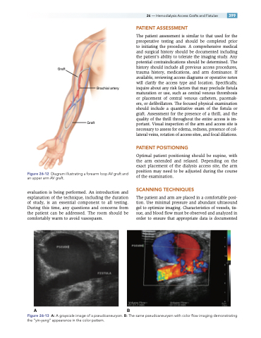
Figure 26-12 Diagram illustrating a forearm loop AV graft and an upper arm AV graft.
evaluation is being performed. An introduction and explanation of the technique, including the duration of study, is an essential component to all testing. During this time, any questions and concerns from the patient can be addressed. The room should be comfortably warm to avoid vasospasm.
AB
Graft
Figure 26-13 A: A grayscale image of a pseudoaneurysm. B: The same pseudoaneurysm with color flow imaging demonstrating the “yin-yang” appearance in the color pattern.
