
Page 415 - Libro 2
P. 415
26 — Hemodialysis Access Grafts and Fistulae
395
Brachial artery
Radial artery
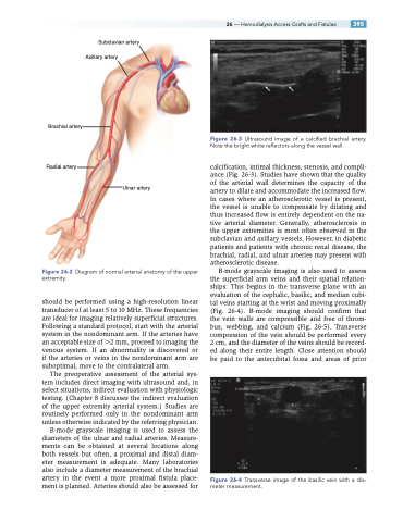
Figure 26-3 Ultrasound image of a calcified brachial artery. Note the bright white reflectors along the vessel wall.
calcification, intimal thickness, stenosis, and compli- ance (Fig. 26-3). Studies have shown that the quality of the arterial wall determines the capacity of the artery to dilate and accommodate the increased flow. In cases where an atherosclerotic vessel is present, the vessel is unable to compensate by dilating and thus increased flow is entirely dependent on the na- tive arterial diameter. Generally, atherosclerosis in the upper extremities is most often observed in the subclavian and axillary vessels. However, in diabetic patients and patients with chronic renal disease, the brachial, radial, and ulnar arteries may present with atherosclerotic disease.
B-mode grayscale imaging is also used to assess the superficial arm veins and their spatial relation- ships. This begins in the transverse plane with an evaluation of the cephalic, basilic, and median cubi- tal veins starting at the wrist and moving proximally (Fig. 26-4). B-mode imaging should confirm that the vein walls are compressible and free of throm- bus, webbing, and calcium (Fig. 26-5). Transverse compression of the vein should be performed every 2 cm, and the diameter of the veins should be record- ed along their entire length. Close attention should be paid to the antecubital fossa and areas of prior
Figure 26-4 Transverse image of the basilic vein with a dia- meter measurement.
Subclavian artery Axillary artery
Figure 26-2 Diagram of normal arterial anatomy of the upper extremity.
should be performed using a high-resolution linear transducer of at least 5 to 10 MHz. These frequencies are ideal for imaging relatively superficial structures. Following a standard protocol, start with the arterial system in the nondominant arm. If the arteries have an acceptable size of 2 mm, proceed to imaging the venous system. If an abnormality is discovered or if the arteries or veins in the nondominant arm are suboptimal, move to the contralateral arm.
The preoperative assessment of the arterial sys- tem includes direct imaging with ultrasound and, in select situations, indirect evaluation with physiologic testing. (Chapter 8 discusses the indirect evaluation of the upper extremity arterial system.) Studies are routinely performed only in the nondominant arm unless otherwise indicated by the referring physician.
B-mode grayscale imaging is used to assess the diameters of the ulnar and radial arteries. Measure- ments can be obtained at several locations along both vessels but often, a proximal and distal diam- eter measurement is adequate. Many laboratories also include a diameter measurement of the brachial artery in the event a more proximal fistula place- ment is planned. Arteries should also be assessed for
Ulnar artery
