
Page 428 - Libro 2
P. 428
408
PART 6 — MISCELLANEOUS
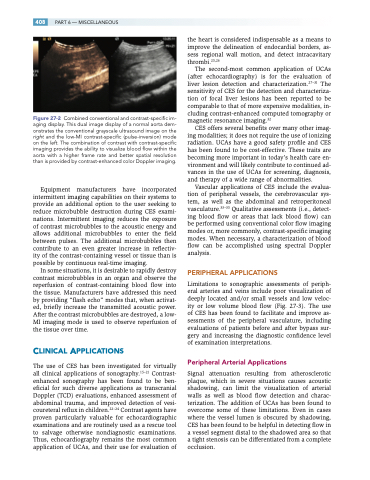
Figure 27-2 Combined conventional and contrast-specific im- aging display. This dual image display of a normal aorta dem- onstrates the conventional grayscale ultrasound image on the right and the low-MI contrast-specific (pulse-inversion) mode on the left. The combination of contrast with contrast-specific imaging provides the ability to visualize blood flow within the aorta with a higher frame rate and better spatial resolution than is provided by contrast-enhanced color Doppler imaging.
Equipment manufacturers have incorporated intermittent imaging capabilities on their systems to provide an additional option to the user seeking to reduce microbubble destruction during CES exami- nations. Intermittent imaging reduces the exposure of contrast microbubbles to the acoustic energy and allows additional microbubbles to enter the field between pulses. The additional microbubbles then contribute to an even greater increase in reflectiv- ity of the contrast-containing vessel or tissue than is possible by continuous real-time imaging.
In some situations, it is desirable to rapidly destroy contrast microbubbles in an organ and observe the reperfusion of contrast-containing blood flow into the tissue. Manufacturers have addressed this need by providing “flash echo” modes that, when activat- ed, briefly increase the transmitted acoustic power. After the contrast microbubbles are destroyed, a low- MI imaging mode is used to observe reperfusion of the tissue over time.
CLINICAL APPLICATIONS
The use of CES has been investigated for virtually all clinical applications of sonography.15–21 Contrast- enhanced sonography has been found to be ben- eficial for such diverse applications as transcranial Doppler (TCD) evaluations, enhanced assessment of abdominal trauma, and improved detection of vesi- coureteral reflux in children.22–24 Contrast agents have proven particularly valuable for echocardiographic examinations and are routinely used as a rescue tool to salvage otherwise nondiagnostic examinations. Thus, echocardiography remains the most common application of UCAs, and their use for evaluation of
the heart is considered indispensable as a means to improve the delineation of endocardial borders, as- sess regional wall motion, and detect intracavitary thrombi.25,26
The second-most common application of UCAs (after echocardiography) is for the evaluation of liver lesion detection and characterization.27–31 The sensitivity of CES for the detection and characteriza- tion of focal liver lesions has been reported to be comparable to that of more expensive modalities, in- cluding contrast-enhanced computed tomography or magnetic resonance imaging.32
CES offers several benefits over many other imag- ing modalities; it does not require the use of ionizing radiation. UCAs have a good safety profile and CES has been found to be cost-effective. These traits are becoming more important in today’s health care en- vironment and will likely contribute to continued ad- vances in the use of UCAs for screening, diagnosis, and therapy of a wide range of abnormalities.
Vascular applications of CES include the evalua- tion of peripheral vessels, the cerebrovascular sys- tem, as well as the abdominal and retroperitoneal vasculature.33–35 Qualitative assessments (i.e., detect- ing blood flow or areas that lack blood flow) can be performed using conventional color flow imaging modes or, more commonly, contrast-specific imaging modes. When necessary, a characterization of blood flow can be accomplished using spectral Doppler analysis.
PERIPHERAL APPLICATIONS
Limitations to sonographic assessments of periph- eral arteries and veins include poor visualization of deeply located and/or small vessels and low veloc- ity or low volume blood flow (Fig. 27-3). The use of CES has been found to facilitate and improve as- sessments of the peripheral vasculature, including evaluations of patients before and after bypass sur- gery and increasing the diagnostic confidence level of examination interpretations.
Peripheral Arterial Applications
Signal attenuation resulting from atherosclerotic plaque, which in severe situations causes acoustic shadowing, can limit the visualization of arterial walls as well as blood flow detection and charac- terization. The addition of UCAs has been found to overcome some of these limitations. Even in cases where the vessel lumen is obscured by shadowing, CES has been found to be helpful in detecting flow in a vessel segment distal to the shadowed area so that a tight stenosis can be differentiated from a complete occlusion.
