
Page 430 - Libro 2
P. 430
410
PART 6 — MISCELLANEOUS
AB
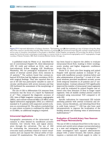
Figure 27-4 Improved delineation of plaque ulceration. The baseline scan (A) demonstrates an area of plaque along the deep wall of the common carotid artery (arrows). After injection of contrast (B), the functional lumen fills with contrast-containing blood, and an area of ulceration (arrowhead) in the plaque is identified. Unlike Doppler sonography, CES provides the ability to directly assess the functional lumen and plaque morphology in a manner similar to conventional angiography and CT angiography. (Image courtesy of Steven Feinstein, MD, Rush University Medical Center, Chicago, IL.)
A published study by Pfister et al. described the use of conventional Doppler US, three-dimensional (3D) US (with and without an UCA), and con- trast-enhanced B-flow imaging (GE Healthcare, Waukesha, WI) for presurgical evaluations of the extent of internal carotid artery (ICA) stenosis in 25 patients.40 The authors found that contrast-en- hanced 3D B-flow had the highest correlation (93%) with surgical findings. They also reported that the use of 3D CES was particularly valuable in cases of circular calcifications with severe stenoses and that CES facilitated the assessment of the morphology of ICA plaque.
The use of CES to differentiate ICA stenoses from occlusions was the focus of a report by Hammond et al.41 They compared the diagnostic accuracy of CES, time-of-flight magnetic resonance angiography (MRA), and contrast-enhanced MRA (CE-MRA) using digital subtraction angiography (DSA) as a reference standard in 31 patients with suspected carotid occlu- sion on conventional US. The authors concluded that in cases where occlusion is confirmed by either CES or CE-MRA, no additional imaging is required.
Intracranial Applications
Sonographic assessments of the intracranial vas- culature is often limited by insufficient acoustic windows, low velocity flow, and signal attenu- ation through the calvarium. The use of CES for evaluating the intracranial circulation can overcome many problems related to vessel visual- ization and can expand the clinical utility of tran- scranial US examinations. The addition of UCAs
has been found to improve the ability to evaluate intracranial blood flow, leading to fewer nondiag- nostic studies and higher diagnostic confidence levels (Fig. 27-6).
Droste et al. used color flow imaging and pulsed Doppler with spectral analysis to evaluate 47 pa- tients with insufficient acoustic windows before and after administration of SonoVue.42 A total of 67 tem- poral windows provided insufficient acoustic access (both temporal windows in 20 patients and 27 uni- lateral studies). Contrast-enhanced TCD significantly improved the number of intracranial vessel segments that could be evaluated by pulsed Doppler and al- lowed color flow detection of flow in longer vessel segments. Only 26 middle cerebral arteries could be evaluated using noncontrast TCD compared to 65 with CES.
Specific indications for transcranial CES include assessing patients with arterial occlusions and ste- noses, venous thrombosis, and detecting blood flow in solid brain tumors.43–45 CES has also been used intraoperatively to improve the localization of the feeding arteries and draining veins of arteriovenous malformations.46
Evaluation of Carotid Artery Vasa Vasorum and Plaque Neovascularity
The use of CES to evaluate blood flow in the vasa va- sorum and carotid artery plaque neovascularization is a new and intriguing area of investigation.47–50 Some investigators have equated atherosclerotic plaque with a tumor because, like tumors, plaque requires a nutrient-rich blood supply in order to
