
Page 432 - Libro 2
P. 432
412
PART 6 — MISCELLANEOUS
Figure 27-6 Contrast-enhanced color Doppler imaging of intracranial vessels. (Image courtesy of Jeff Powers, PhD, Philips Ultrasound, bothell, WA)
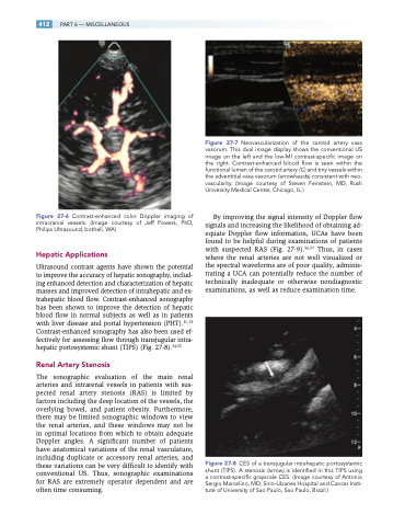
Hepatic Applications
Ultrasound contrast agents have shown the potential to improve the accuracy of hepatic sonography, includ- ing enhanced detection and characterization of hepatic masses and improved detection of intrahepatic and ex- trahepatic blood flow. Contrast-enhanced sonography has been shown to improve the detection of hepatic blood flow in normal subjects as well as in patients with liver disease and portal hypertension (PHT).51–53 Contrast-enhanced sonography has also been used ef- fectively for assessing flow through transjugular intra- hepatic portosystemic shunt (TIPS) (Fig. 27-8).54,55
Renal Artery Stenosis
The sonographic evaluation of the main renal arteries and intrarenal vessels in patients with sus- pected renal artery stenosis (RAS) is limited by factors including the deep location of the vessels, the overlying bowel, and patient obesity. Furthermore, there may be limited sonographic windows to view the renal arteries, and these windows may not be in optimal locations from which to obtain adequate Doppler angles. A significant number of patients have anatomical variations of the renal vasculature, including duplicate or accessory renal arteries, and these variations can be very difficult to identify with conventional US. Thus, sonographic examinations for RAS are extremely operator dependent and are often time consuming.
Figure 27-7 Neovascularization of the carotid artery vasa vasorum. This dual image display shows the conventional US image on the left and the low-MI contrast-specific image on the right. Contrast-enhanced blood flow is seen within the functional lumen of the carotid artery (C) and tiny vessels within the adventitial vasa vasorum (arrowheads) consistent with neo- vascularity. (Image courtesy of Steven Feinstein, MD, Rush University Medical Center, Chicago, IL.)
By improving the signal intensity of Doppler flow signals and increasing the likelihood of obtaining ad- equate Doppler flow information, UCAs have been found to be helpful during examinations of patients with suspected RAS (Fig. 27-9).56,57 Thus, in cases where the renal arteries are not well visualized or the spectral waveforms are of poor quality, adminis- trating a UCA can potentially reduce the number of technically inadequate or otherwise nondiagnostic examinations, as well as reduce examination time.
Figure 27-8 CES of a transjugular intrahepatic portosystemic shunt (TIPS). A stenosis (arrow) is identified in this TIPS using a contrast-specific grayscale CES. (Image courtesy of Antonio Sergio Marcelino, MD, Sirio-Libanes Hospital and Cancer Insti- tute of University of Sao Paulo, Sao Paulo, Brazil.)
