
Page 434 - Libro 2
P. 434
414
PART 6 — MISCELLANEOUS
AB
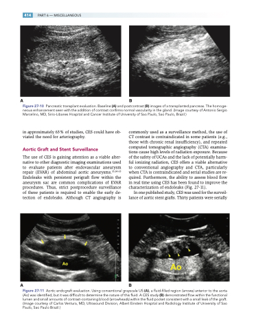
Figure 27-10 Pancreatic transplant evaluation. Baseline (A) and postcontrast (B) images of a transplanted pancreas. The homoge- neous enhancement seen with the addition of contrast confirms normal vascularity in the gland. (Image courtesy of Antonio Sergio Marcelino, MD, Sirio-Libanes Hospital and Cancer Institute of University of Sao Paulo, Sao Paulo, Brazil.)
in approximately 63% of studies, CES could have ob- viated the need for arteriography.
Aortic Graft and Stent Surveillance
The use of CES is gaining attention as a viable alter- native to other diagnostic imaging examinations used to evaluate patients after endovascular aneurysm repair (EVAR) of abdominal aortic aneurysms.35,64-65 Endoleaks with persistent perigraft flow within the aneurysm sac are common complications of EVAR procedures. Thus, strict postprocedure surveillance of these patients is required to enable the early de- tection of endoleaks. Although CT angiography is
commonly used as a surveillance method, the use of CT contrast is contraindicated in some patients (e.g., those with chronic renal insufficiency), and repeated computed tomographic angiography (CTA) examina- tions cause high levels of radiation exposure. Because of the safety of UCAs and the lack of potentially harm- ful ionizing radiation, CES offers a viable alternative to conventional angiography and CTA, particularly when CTA is contraindicated and serial studies are re- quired. Furthermore, the ability to assess blood flow in real time using CES has been found to improve the characterization of endoleaks (Fig. 27-11).
In one published study, CES was used for the surveil- lance of aortic stent grafts. Thirty patients were serially
AB
Figure 27-11 Aortic endograft evaluation. Using conventional grayscale US (A), a fluid-filled region (arrows) anterior to the aorta (Ao) was identified, but it was difficult to determine the nature of this fluid. A CES study (B) demonstrated flow within the functional lumen and small amounts of contrast-containing blood (arrowheads) within the fluid pocket consistent with a small leak of the graft. (Image courtesy of Carlos Ventura, MD, Ultrasound Division, Albert Einstein Hospital and Radiology Institute of University of Sao Paulo, Sao Paulo Brazil.)
