
Page 435 - Libro 2
P. 435
27 — Vascular Applications of Ultrasound Contrast Agents
415
evaluated with CES, and the results were compared to either CTA or magnetic resonance angiography (MRA) as the gold standard. All CTA/MRA-detected endoleaks were detected by CES, yielding a 100% sensitivity.
Other Abdominal Applications
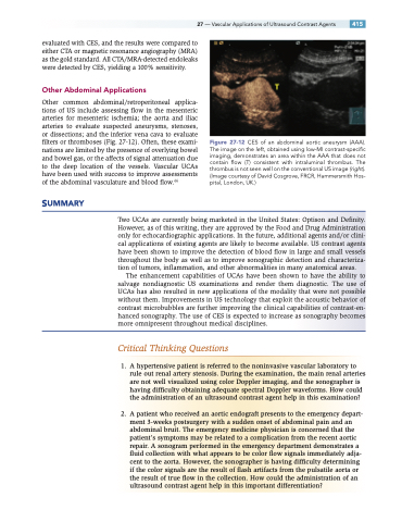
Other common abdominal/retroperitoneal applica- tions of US include assessing flow in the mesenteric arteries for mesenteric ischemia; the aorta and iliac arteries to evaluate suspected aneurysms, stenoses, or dissections; and the inferior vena cava to evaluate filters or thromboses (Fig. 27-12). Often, these exami- nations are limited by the presence of overlying bowel and bowel gas, or the affects of signal attenuation due to the deep location of the vessels. Vascular UCAs have been used with success to improve assessments of the abdominal vasculature and blood flow.66
Figure 27-12 CES of an abdominal aortic aneurysm (AAA). The image on the left, obtained using low-MI contrast-specific imaging, demonstrates an area within the AAA that does not contain flow (T) consistent with intraluminal thrombus. The thrombus is not seen well on the conventional US image (right). (Image courtesy of David Cosgrove, FRCR, Hammersmith Hos- pital, London, UK.)
SUMMARY
Two UCAs are currently being marketed in the United States: Optison and Definity. However, as of this writing, they are approved by the Food and Drug Administration only for echocardiographic applications. In the future, additional agents and/or clini- cal applications of existing agents are likely to become available. US contrast agents have been shown to improve the detection of blood flow in large and small vessels throughout the body as well as to improve sonographic detection and characteriza- tion of tumors, inflammation, and other abnormalities in many anatomical areas.
The enhancement capabilities of UCAs have been shown to have the ability to salvage nondiagnostic US examinations and render them diagnostic. The use of UCAs has also resulted in new applications of the modality that were not possible without them. Improvements in US technology that exploit the acoustic behavior of contrast microbubbles are further improving the clinical capabilities of contrast-en- hanced sonography. The use of CES is expected to increase as sonography becomes more omnipresent throughout medical disciplines.
Critical Thinking Questions
1. A hypertensive patient is referred to the noninvasive vascular laboratory to rule out renal artery stenosis. During the examination, the main renal arteries are not well visualized using color Doppler imaging, and the sonographer is having difficulty obtaining adequate spectral Doppler waveforms. How could the administration of an ultrasound contrast agent help in this examination?
2. A patient who received an aortic endograft presents to the emergency depart- ment 3-weeks postsurgery with a sudden onset of abdominal pain and an abdominal bruit. The emergency medicine physician is concerned that the patient’s symptoms may be related to a complication from the recent aortic repair. A sonogram performed in the emergency department demonstrates a fluid collection with what appears to be color flow signals immediately adja- cent to the aorta. However, the sonographer is having difficulty determining if the color signals are the result of flash artifacts from the pulsatile aorta or the result of true flow in the collection. How could the administration of an ultrasound contrast agent help in this important differentiation?
