
Page 429 - Libro 2
P. 429
27 — Vascular Applications of Ultrasound Contrast Agents
409
AB
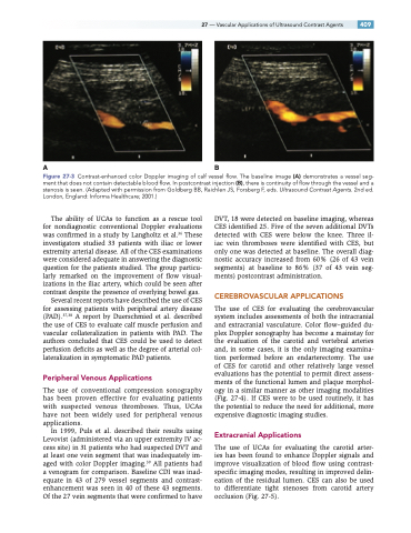
Figure 27-3 Contrast-enhanced color Doppler imaging of calf vessel flow. The baseline image (A) demonstrates a vessel seg- ment that does not contain detectable blood flow. In postcontrast injection (B), there is continuity of flow through the vessel and a stenosis is seen. (Adapted with permission from Goldberg BB, Raichlen JS, Forsberg F, eds. Ultrasound Contrast Agents. 2nd ed. London, England: Informa Healthcare; 2001.)
The ability of UCAs to function as a rescue tool for nondiagnostic conventional Doppler evaluations was confirmed in a study by Langholtz et al.36 These investigators studied 33 patients with iliac or lower extremity arterial disease. All of the CES examinations were considered adequate in answering the diagnostic question for the patients studied. The group particu- larly remarked on the improvement of flow visual- izations in the iliac artery, which could be seen after contrast despite the presence of overlying bowel gas.
Several recent reports have described the use of CES for assessing patients with peripheral artery disease (PAD).37,38 A report by Duerschmied et al. described the use of CES to evaluate calf muscle perfusion and vascular collateralization in patients with PAD. The authors concluded that CES could be used to detect perfusion deficits as well as the degree of arterial col- lateralization in symptomatic PAD patients.
Peripheral Venous Applications
The use of conventional compression sonography has been proven effective for evaluating patients with suspected venous thromboses. Thus, UCAs have not been widely used for peripheral venous applications.
In 1999, Puls et al. described their results using Levovist (administered via an upper extremity IV ac- cess site) in 31 patients who had suspected DVT and at least one vein segment that was inadequately im- aged with color Doppler imaging.39 All patients had a venogram for comparison. Baseline CDI was inad- equate in 43 of 279 vessel segments and contrast- enhancement was seen in 40 of these 43 segments. Of the 27 vein segments that were confirmed to have
DVT, 18 were detected on baseline imaging, whereas CES identified 25. Five of the seven additional DVTs detected with CES were below the knee. Three il- iac vein thromboses were identified with CES, but only one was detected at baseline. The overall diag- nostic accuracy increased from 60% (26 of 43 vein segments) at baseline to 86% (37 of 43 vein seg- ments) postcontrast administration.
CEREBROVASCULAR APPLICATIONS
The use of CES for evaluating the cerebrovascular system includes assessments of both the intracranial and extracranial vasculature. Color flow–guided du- plex Doppler sonography has become a mainstay for the evaluation of the carotid and vertebral arteries and, in some cases, it is the only imaging examina- tion performed before an endarterectomy. The use of CES for carotid and other relatively large vessel evaluations has the potential to permit direct assess- ments of the functional lumen and plaque morphol- ogy in a similar manner as other imaging modalities (Fig. 27-4). If CES were to be used routinely, it has the potential to reduce the need for additional, more expensive diagnostic imaging studies.
Extracranial Applications
The use of UCAs for evaluating the carotid arter- ies has been found to enhance Doppler signals and improve visualization of blood flow using contrast- specific imaging modes, resulting in improved delin- eation of the residual lumen. CES can also be used to differentiate tight stenoses from carotid artery occlusion (Fig. 27-5).
