
Page 57 - Libro 2
P. 57
4 — The Extracranial Duplex Ultrasound Examination 37
TABLE 4-1
Patient Interview for Pertinent Medical History
• Are you being treated for high blood pressure (hypertension)?
• Are you being treated for high cholesterol (hypercholesterolemia)?
• Do you have diabetes? If so, for how long? Is it treated with diet control, oral medication, or insulin?
• Have you had a heart attack (myocardial infarction) or chest pains?
• Have you ever had surgery or other interventions on any of your blood vessels (coronary artery bypass graft or stent placement, carotid endarterectomy
or stent placement, peripheral arterial revascularization)?
• Have you had a stroke or “mini stroke” (CVA, TIA) in the past?
• Have you recently or are you currently experiencing stroke-like symptoms that include:
o Weakness or numbness down one side of your
body
o Difficulty with balance or walking
o Slurred speech or difficulty forming words
o Dizziness, nausea, or vomiting
o Severeheadache
o Vision disturbances such as “cloudiness” or perhaps like a “shade” coming down over one eye
with a stethoscope for bruits (high, middle, and low neck, and in the region of the clavicle).
PATIENT POSITIONING
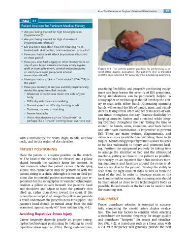
Place the patient in a supine position on the stretch- er. The head of the bed may be elevated and a pillow placed beneath the patient’s knees for comfort. In rare instances when the patient cannot tolerate lying supine, this examination may be performed with the patient sitting in a chair, although it is not an ideal po- sition due to potential patient movement and poor er- gonomics for the sonographer or vascular technologist. Position a pillow equally beneath the patient’s head and shoulders and adjust to have the patient’s chin tilted up, rather than down toward the chest. If this cannot be accomplished, forego the pillow and provide a towel underneath the patient’s neck for support. The patient’s head should be turned away from the side examined, approximately 45° from midline (Fig. 4-1).
Avoiding Repetitive Stress Injury
Career longevity depends greatly on proper sonog- rapher/technologist positioning by helping to avoid repetitive stress injuries (RSIs). Being ambidextrous,
Figure 4-1 The correct patient position for performing a ca- rotid artery duplex evaluation. The patient’s chin is elevated and the head is turned 45° away from the side being examined.
practicing flexibility, and properly positioning equip- ment can help lessen the severity of RSI symptoms. Being ambidextrous can be particularly helpful. A sonographer or technologist should develop the abil- ity to scan with either hand. Alternating scanning hands will extend the life of hands, arms, and shoul- ders by taking stress off of one set of muscles at vari- ous times throughout the day. Practice flexibility by keeping muscles limber and stretched while keep- ing hydrated throughout the day. Taking the time to stretch the hands, arms, shoulders, and back before and after each examination is imperative to prevent RSI. There are many written, diagrammatic, and video resources available demonstrating these tech- niques. Maintaining proper hydration allows muscles to be less vulnerable to injury and promotes heal- ing. Position the equipment properly by taking time to arrange the stretcher or bed and the ultrasound machine, getting as close to the patient as possible. Particularly on an inpatient floor, this involves mov- ing equipment and furniture around the room to al- low access close to the patient. Develop the ability to scan from the right and left sides as well as from the head of the bed. In order to decrease strain on the neck and shoulder muscles, the scanning arm should be maintained as close to the technologist’s body as possible. Rolled towels or the bed can be used to rest the scanning arm.
EQUIPMENT
Proper transducer selection is essential to success- fully completing the carotid artery duplex evalua- tion. The two key factors to consider when selecting a transducer are transmit frequency for image quality and transducer “footprint” for access and visualiza- tion (Fig. 4-2). A transducer such as a linear array with a 7-4 MHz frequency will generally provide the best
