
Page 58 - Libro 2
P. 58
38
PART 2 — CEREBROVASCULAR
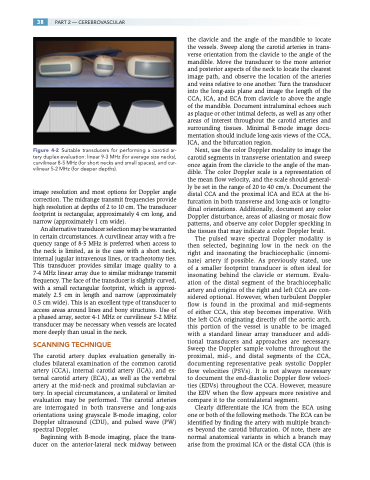
Figure 4-2 Suitable transducers for performing a carotid ar- tery duplex evaluation: linear 9-3 MHz (for average size necks), curvilinear 8-5 MHz (for short necks and small spaces), and cur- vilinear 5-2 MHz (for deeper depths).
image resolution and most options for Doppler angle correction. The midrange transmit frequencies provide high resolution at depths of 2 to 10 cm. The transducer footprint is rectangular, approximately 4 cm long, and narrow (approximately 1 cm wide).
An alternative transducer selection may be warranted in certain circumstances. A curvilinear array with a fre- quency range of 8-5 MHz is preferred when access to the neck is limited, as is the case with a short neck, internal jugular intravenous lines, or tracheotomy ties. This transducer provides similar image quality to a 7-4 MHz linear array due to similar midrange transmit frequency. The face of the transducer is slightly curved, with a small rectangular footprint, which is approxi- mately 2.5 cm in length and narrow (approximately 0.5 cm wide). This is an excellent type of transducer to access areas around lines and bony structures. Use of a phased array, sector 4-1 MHz or curvilinear 5-2 MHz transducer may be necessary when vessels are located more deeply than usual in the neck.
SCANNING TECHNIQUE
The carotid artery duplex evaluation generally in- cludes bilateral examination of the common carotid artery (CCA), internal carotid artery (ICA), and ex- ternal carotid artery (ECA), as well as the vertebral artery at the mid-neck and proximal subclavian ar- tery. In special circumstances, a unilateral or limited evaluation may be performed. The carotid arteries are interrogated in both transverse and long-axis orientations using grayscale B-mode imaging, color Doppler ultrasound (CDU), and pulsed wave (PW) spectral Doppler.
Beginning with B-mode imaging, place the trans- ducer on the anterior-lateral neck midway between
the clavicle and the angle of the mandible to locate the vessels. Sweep along the carotid arteries in trans- verse orientation from the clavicle to the angle of the mandible. Move the transducer to the more anterior and posterior aspects of the neck to locate the clearest image path, and observe the location of the arteries and veins relative to one another. Turn the transducer into the long-axis plane and image the length of the CCA, ICA, and ECA from clavicle to above the angle of the mandible. Document intraluminal echoes such as plaque or other intimal defects, as well as any other areas of interest throughout the carotid arteries and surrounding tissues. Minimal B-mode image docu- mentation should include long-axis views of the CCA, ICA, and the bifurcation region.
Next, use the color Doppler modality to image the carotid segments in transverse orientation and sweep once again from the clavicle to the angle of the man- dible. The color Doppler scale is a representation of the mean flow velocity, and the scale should general- ly be set in the range of 20 to 40 cm/s. Document the distal CCA and the proximal ICA and ECA at the bi- furcation in both transverse and long-axis or longitu- dinal orientations. Additionally, document any color Doppler disturbance, areas of aliasing or mosaic flow patterns, and observe any color Doppler speckling in the tissues that may indicate a color Doppler bruit.
The pulsed wave spectral Doppler modality is then selected, beginning low in the neck on the right and insonating the brachiocephalic (innomi- nate) artery if possible. As previously stated, use of a smaller footprint transducer is often ideal for insonating behind the clavicle or sternum. Evalu- ation of the distal segment of the brachiocephalic artery and origins of the right and left CCA are con- sidered optional. However, when turbulent Doppler flow is found in the proximal and mid-segments of either CCA, this step becomes imperative. With the left CCA originating directly off the aortic arch, this portion of the vessel is unable to be imaged with a standard linear array transducer and addi- tional transducers and approaches are necessary. Sweep the Doppler sample volume throughout the proximal, mid-, and distal segments of the CCA, documenting representative peak systolic Doppler flow velocities (PSVs). It is not always necessary to document the end-diastolic Doppler flow veloci- ties (EDVs) throughout the CCA. However, measure the EDV when the flow appears more resistive and compare it to the contralateral segment.
Clearly differentiate the ICA from the ECA using one or both of the following methods. The ECA can be identified by finding the artery with multiple branch- es beyond the carotid bifurcation. Of note, there are normal anatomical variants in which a branch may arise from the proximal ICA or the distal CCA (this is
