
Page 60 - Libro 2
P. 60
40 PART 2 — CEREBROVASCULAR
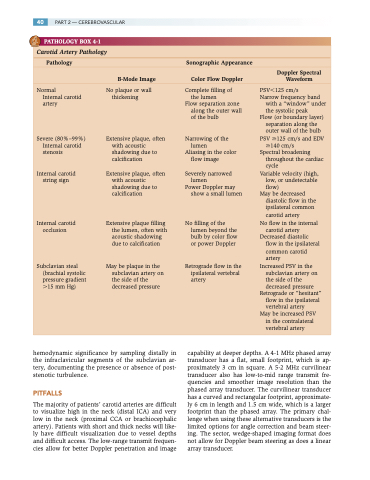
PATHOLOGY BOX 4-1
Carotid Artery Pathology
Pathology Sonographic Appearance
Doppler Spectral B-Mode Image Color Flow Doppler Waveform
Normal
Internal carotid artery
Severe (80%–99%) Internal carotid stenosis
Internal carotid string sign
Internal carotid occlusion
Subclavian steal (brachial systolic pressure gradient 15 mm Hg)
No plaque or wall thickening
Extensive plaque, often with acoustic shadowing due to calcification
Extensive plaque, often with acoustic shadowing due to calcification
Extensive plaque filling the lumen, often with acoustic shadowing due to calcification
May be plaque in the subclavian artery on the side of the decreased pressure
Complete filling of the lumen
Flow separation zone along the outer wall of the bulb
Narrowing of the lumen
Aliasing in the color flow image
Severely narrowed lumen
Power Doppler may show a small lumen
No filling of the lumen beyond the bulb by color flow or power Doppler
Retrograde flow in the ipsilateral vertebral artery
PSV125 cm/s
Narrow frequency band
with a “window” under
the systolic peak
Flow (or boundary layer)
separation along the
outer wall of the bulb PSV 125 cm/s and EDV
140 cm/s Spectral broadening
throughout the cardiac
cycle
Variable velocity (high,
low, or undetectable
flow)
May be decreased
diastolic flow in the ipsilateral common carotid artery
No flow in the internal carotid artery
Decreased diastolic flow in the ipsilateral common carotid artery
Increased PSV in the subclavian artery on the side of the decreased pressure
Retrograde or “hesitant” flow in the ipsilateral vertebral artery
May be increased PSV
in the contralateral vertebral artery
hemodynamic significance by sampling distally in the infraclavicular segments of the subclavian ar- tery, documenting the presence or absence of post- stenotic turbulence.
PITFALLS
The majority of patients’ carotid arteries are difficult to visualize high in the neck (distal ICA) and very low in the neck (proximal CCA or brachiocephalic artery). Patients with short and thick necks will like- ly have difficult visualization due to vessel depths and difficult access. The low-range transmit frequen- cies allow for better Doppler penetration and image
capability at deeper depths. A 4-1 MHz phased array transducer has a flat, small footprint, which is ap- proximately 3 cm in square. A 5-2 MHz curvilinear transducer also has low-to-mid range transmit fre- quencies and smoother image resolution than the phased array transducer. The curvilinear transducer has a curved and rectangular footprint, approximate- ly 6 cm in length and 1.5 cm wide, which is a larger footprint than the phased array. The primary chal- lenge when using these alternative transducers is the limited options for angle correction and beam steer- ing. The sector, wedge-shaped imaging format does not allow for Doppler beam steering as does a linear array transducer.
