
Page 62 - Libro 2
P. 62
42
PART 2 — CEREBROVASCULAR
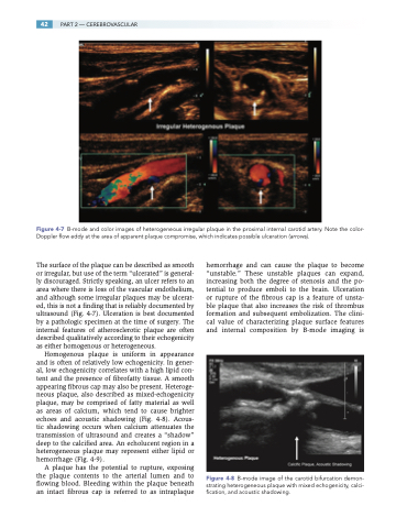
Figure 4-7 B-mode and color images of heterogeneous irregular plaque in the proximal internal carotid artery. Note the color- Doppler flow eddy at the area of apparent plaque compromise, which indicates possible ulceration (arrows).
The surface of the plaque can be described as smooth or irregular, but use of the term “ulcerated” is general- ly discouraged. Strictly speaking, an ulcer refers to an area where there is loss of the vascular endothelium, and although some irregular plaques may be ulcerat- ed, this is not a finding that is reliably documented by ultrasound (Fig. 4-7). Ulceration is best documented by a pathologic specimen at the time of surgery. The internal features of atherosclerotic plaque are often described qualitatively according to their echogenicity as either homogenous or heterogeneous.
Homogenous plaque is uniform in appearance and is often of relatively low echogenicity. In gener- al, low echogenicity correlates with a high lipid con- tent and the presence of fibrofatty tissue. A smooth appearing fibrous cap may also be present. Heteroge- neous plaque, also described as mixed-echogenicity plaque, may be comprised of fatty material as well as areas of calcium, which tend to cause brighter echoes and acoustic shadowing (Fig. 4-8). Acous- tic shadowing occurs when calcium attenuates the transmission of ultrasound and creates a “shadow” deep to the calcified area. An echolucent region in a heterogeneous plaque may represent either lipid or hemorrhage (Fig. 4-9).
A plaque has the potential to rupture, exposing the plaque contents to the arterial lumen and to flowing blood. Bleeding within the plaque beneath an intact fibrous cap is referred to as intraplaque
hemorrhage and can cause the plaque to become “unstable.” These unstable plaques can expand, increasing both the degree of stenosis and the po- tential to produce emboli to the brain. Ulceration or rupture of the fibrous cap is a feature of unsta- ble plaque that also increases the risk of thrombus formation and subsequent embolization. The clini- cal value of characterizing plaque surface features and internal composition by B-mode imaging is
Figure 4-8 B-mode image of the carotid bifurcation demon- strating heterogeneous plaque with mixed echogenicity, calci- fication, and acoustic shadowing.
