
Page 61 - Libro 2
P. 61
4 — The Extracranial Duplex Ultrasound Examination
41
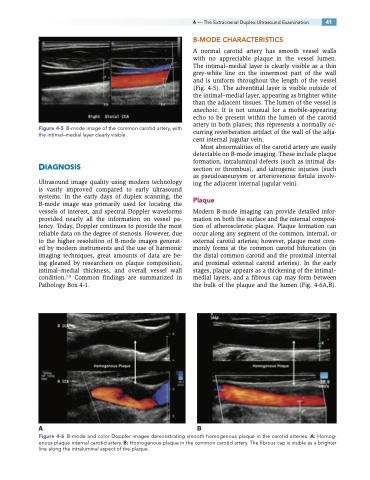
Figure 4-5 B-mode image of the common carotid artery, with the intimal–medial layer clearly visible.
DIAGNOSIS
Ultrasound image quality using modern technology is vastly improved compared to early ultrasound systems. In the early days of duplex scanning, the B-mode image was primarily used for locating the vessels of interest, and spectral Doppler waveforms provided nearly all the information on vessel pa- tency. Today, Doppler continues to provide the most reliable data on the degree of stenosis. However, due to the higher resolution of B-mode images generat- ed by modern instruments and the use of harmonic imaging techniques, great amounts of data are be- ing gleaned by researchers on plaque composition, intimal–medial thickness, and overall vessel wall condition.7,8 Common findings are summarized in Pathology Box 4-1.
B-MODE CHARACTERISTICS
A normal carotid artery has smooth vessel walls with no appreciable plaque in the vessel lumen. The intimal–medial layer is clearly visible as a thin grey-white line on the innermost part of the wall and is uniform throughout the length of the vessel (Fig. 4-5). The adventitial layer is visible outside of the intimal–medial layer, appearing as brighter white than the adjacent tissues. The lumen of the vessel is anechoic. It is not unusual for a mobile-appearing echo to be present within the lumen of the carotid artery in both planes; this represents a normally oc- curring reverberation artifact of the wall of the adja- cent internal jugular vein.
Most abnormalities of the carotid artery are easily detectable on B-mode imaging. These include plaque formation, intraluminal defects (such as intimal dis- section or thrombus), and iatrogenic injuries (such as pseudoaneurysm or arteriovenous fistula involv- ing the adjacent internal jugular vein).
Plaque
Modern B-mode imaging can provide detailed infor- mation on both the surface and the internal composi- tion of atherosclerotic plaque. Plaque formation can occur along any segment of the common, internal, or external carotid arteries; however, plaque most com- monly forms at the common carotid bifurcation (in the distal common carotid and the proximal internal and proximal external carotid arteries). In the early stages, plaque appears as a thickening of the intimal– medial layers, and a fibrous cap may form between the bulk of the plaque and the lumen (Fig. 4-6A,B).
AB
Figure 4-6 B-mode and color Doppler images demonstrating smooth homogenous plaque in the carotid arteries. A: Homog- enous plaque internal carotid artery. B: Homogenous plaque in the common carotid artery. The fibrous cap is visible as a brighter line along the intraluminal aspect of the plaque.
