
Page 59 - Libro 2
P. 59
4 — The Extracranial Duplex Ultrasound Examination
39
Figure 4-3 Duplex image of the external carotid artery. The Doppler flow signal is affected by oscillations on the ipsilateral temporal artery.
generally the superior thyroid artery). A second tech- nique involves the palpation of the pulse of the su- perficial temporal artery just anterior to the ear, then oscillating the flow by “tapping” on the artery (the “temporal tap” maneuver) while insonating flow in the proximal segment of the ECA (Fig. 4-3). An arti- fact from the tapping (oscillations) should be visible in the spectral waveform from the ECA but not from the ICA. The temporal tap technique is not always accurate, particularly in patients with an ICA occlu- sion and an ECA collateralization. In some patients, the temporal tap may also produce oscillations within the ICA if the temporal artery is tapped too strong- ly. In addition, if the temporal artery is inadequately tapped, oscillations will not be generated within the ECA Doppler spectrum. Therefore, the temporal tap can be helpful in many patients but should be used with caution.
Sweep the Doppler sample volume from the distal CCA into the ECA, continuously insonating to document the highest peak systolic velocity in the proximal ECA segment. Continue to move the Doppler sample volume through the proximal and mid-segments to determine whether any waveform changes are present. When elevated flow velocities are obtained in the ECA, document the presence or absence of poststenotic turbulence to help determine hemodynamic significance.
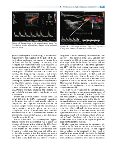
Return to the distal CCA and sweep the Doppler sample volume into the proximal ICA to detect the presence of stenosis at the origin of the ICA. Docu- ment flow separation in the carotid bulb, if present, which appears as a small area of flow reversal. This typically is located along the outer wall of the bulb on the side opposite to the flow divider, as shown in Figure 4-4. As plaque formation progresses and fills in the carotid bulb, the area of flow separation
Figure 4-4 Duplex image of normal Doppler flow separation in the proximal internal carotid artery (carotid bulb).
disappears. It is not necessary to measure the flow velocity of this reversal component. Laminar flow may actually be difficult to demonstrate in patients with large carotid bulbs. Move the sample volume throughout the carotid bulb to detect the highest PSV and EDV with the most laminar waveform obtain- able. Continue to insonate through the carotid bulb and the proximal, mid-, and distal segments of the ICA. Often, the distal segment of the ICA is difficult to visualize or insonate beyond the angle of the man- dible. It is particularly important to evaluate these segments of the ICA in patients who are at risk for fibromuscular dysplasia (young- to middle-aged fe- males). For this application, curved or phased array transducers are ideal.
The next vessel evaluated is the vertebral artery. Place the transducer at the anterior-medial aspect of the mid-neck in the long-axis position. Once the CCA has been identified, slowly slide or angle the trans- ducer posteriorly, focusing deep to the CCA to view the vertebral artery between the transverse processes of the cervical vertebrae. Take care to properly iden- tify flow direction and document waveform contour at the level of the mid-neck. An abnormal waveform contour or flow direction indicates hemodynamically significant stenosis of the ipsilateral proximal subcla- vian artery, which will be discussed later. When tur- bulent Doppler flow is detected in the vertebral artery at low-to-mid neck, evaluate the origin and proximal segments to identify a stenosis.
Finally, place the transducer in transverse ori- entation at the base of the neck to insonate the supraclavicular subclavian artery. Set the Doppler sample volume as far proximally in the vessel as possible, then sweep distally to obtain the highest PSV and the most laminar Doppler spectral wave- form. If elevated velocities are detected, determine
