
Page 63 - Libro 2
P. 63
4 — The Extracranial Duplex Ultrasound Examination
43
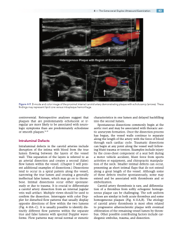
Figure 4-9 B-mode and color image of the proximal internal carotid artery demonstrating plaque with echolucency (arrows). These findings may represent lipid core versus intraplaque hemorrhage.
controversial. Retrospective analyses suggest that plaques that are predominately echolucent or ir- regular are more likely to be associated with neuro- logic symptoms than are predominately echodense or smooth plaques.8–10
Intraluminal Defects
Intraluminal defects in the carotid arteries include disruption of the intima with blood from the true lumen flowing between the layers of the vessel wall. This separation of the layers is referred to as an arterial dissection and creates a second (false) flow lumen within the vessel. (Chapter 5 will pres- ent additional examples of dissections.) Dissections tend to occur in a spiral pattern along the vessel, narrowing the true lumen and creating a generally ineffectual false lumen, which may go on to throm- bose. Intimal dissections occur either spontane- ously or due to trauma. It is crucial to differentiate a carotid artery dissection from an internal jugular vein wall artifact. Multiple views should be used to confirm the dissection. Interrogate with color Dop- pler for disturbed flow patterns that usually display opposite directions of flow within the two lumens (Fig. 4-10A–C). It is usually possible to identify dis- tinctly different flow patterns within the separate true and false lumens with spectral Doppler wave- forms. Flow patterns may reveal normal or stenotic
characteristics in one lumen and delayed backfilling into the second lumen.
Spontaneous dissections commonly begin at the aortic root and may be associated with thoracic aor- tic aneurysm formation. Once the dissection process has begun, the vessel walls continue to separate along the length of the artery with the force of blood through each cardiac cycle. Traumatic dissections can begin at any point along the vessel wall follow- ing blunt trauma or torsion. Examples include injury by the cross-chest component of a seat belt during a motor vehicle accident, blunt force from sports activities or equipment, and chiropractic manipula- tion of the neck. Smaller intimal defects can occur, presenting as short intimal flaps that do not extend along a great length of the vessel. Although some of these defects resolve spontaneously, some may extend and be associated with thrombosis and or embolization.
Carotid artery thrombosis is rare, and differentia- tion of a thrombus from softly echogenic homoge- neous plaque can be challenging. The soft uniform echoes are similar in both acute thrombus and some homogeneous plaques (Fig. 4-11A,B). The etiology of carotid artery thrombosis is most often related to progressive atherosclerotic plaque with eventual obliteration of the remaining vessel lumen by throm- bus. Other possible contributing factors include car- diogenic embolus, trauma, and dissection.
