
Page 65 - Libro 2
P. 65
4 — The Extracranial Duplex Ultrasound Examination
45
AB
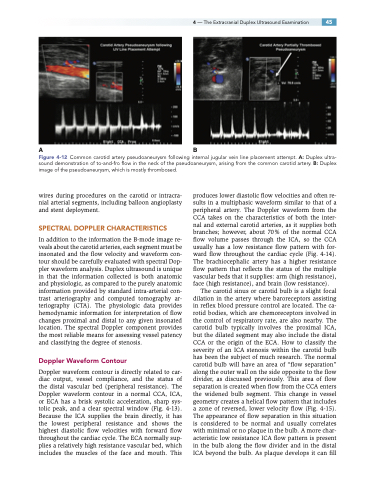
Figure 4-12 Common carotid artery pseudoaneurysm following internal jugular vein line placement attempt. A: Duplex ultra- sound demonstration of to-and-fro flow in the neck of the pseudoaneurysm, arising from the common carotid artery. B: Duplex image of the pseudoaneurysm, which is mostly thrombosed.
wires during procedures on the carotid or intracra- nial arterial segments, including balloon angioplasty and stent deployment.
SPECTRAL DOPPLER CHARACTERISTICS
In addition to the information the B-mode image re- veals about the carotid arteries, each segment must be insonated and the flow velocity and waveform con- tour should be carefully evaluated with spectral Dop- pler waveform analysis. Duplex ultrasound is unique in that the information collected is both anatomic and physiologic, as compared to the purely anatomic information provided by standard intra-arterial con- trast arteriography and computed tomography ar- teriography (CTA). The physiologic data provides hemodynamic information for interpretation of flow changes proximal and distal to any given insonated location. The spectral Doppler component provides the most reliable means for assessing vessel patency and classifying the degree of stenosis.
Doppler Waveform Contour
Doppler waveform contour is directly related to car- diac output, vessel compliance, and the status of the distal vascular bed (peripheral resistance). The Doppler waveform contour in a normal CCA, ICA, or ECA has a brisk systolic acceleration, sharp sys- tolic peak, and a clear spectral window (Fig. 4-13). Because the ICA supplies the brain directly, it has the lowest peripheral resistance and shows the highest diastolic flow velocities with forward flow throughout the cardiac cycle. The ECA normally sup- plies a relatively high resistance vascular bed, which includes the muscles of the face and mouth. This
produces lower diastolic flow velocities and often re- sults in a multiphasic waveform similar to that of a peripheral artery. The Doppler waveform from the CCA takes on the characteristics of both the inter- nal and external carotid arteries, as it supplies both branches; however, about 70% of the normal CCA flow volume passes through the ICA, so the CCA usually has a low resistance flow pattern with for- ward flow throughout the cardiac cycle (Fig. 4-14). The brachiocephalic artery has a higher resistance flow pattern that reflects the status of the multiple vascular beds that it supplies: arm (high resistance), face (high resistance), and brain (low resistance).
The carotid sinus or carotid bulb is a slight focal dilation in the artery where baroreceptors assisting in reflex blood pressure control are located. The ca- rotid bodies, which are chemoreceptors involved in the control of respiratory rate, are also nearby. The carotid bulb typically involves the proximal ICA, but the dilated segment may also include the distal CCA or the origin of the ECA. How to classify the severity of an ICA stenosis within the carotid bulb has been the subject of much research. The normal carotid bulb will have an area of “flow separation” along the outer wall on the side opposite to the flow divider, as discussed previously. This area of flow separation is created when flow from the CCA enters the widened bulb segment. This change in vessel geometry creates a helical flow pattern that includes a zone of reversed, lower velocity flow (Fig. 4-15). The appearance of flow separation in this situation is considered to be normal and usually correlates with minimal or no plaque in the bulb. A more char- acteristic low resistance ICA flow pattern is present in the bulb along the flow divider and in the distal ICA beyond the bulb. As plaque develops it can fill
