
Page 67 - Libro 2
P. 67
4 — The Extracranial Duplex Ultrasound Examination
47
Figure 4-15 Duplex image of normal Doppler arterial flow characteristics through the carotid bulb and internal carotid artery.
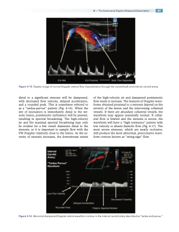
distal to a significant stenosis will be dampened, with decreased flow velocity, delayed acceleration, and a rounded peak. This is sometimes referred to as a “tardus-parvus” pattern (Fig. 4-16). When the site of insonation is immediately distal to the ste- notic lesion, poststenotic turbulence will be present, resulting in spectral broadening. The high-velocity jet and the maximal spectral broadening may only be evident for a few vessel diameters distal to the stenosis, so it is important to sample flow with the PW Doppler relatively close to the lesion. As the se- verity of stenosis increases, the downstream extent
of the high-velocity jet and dampened poststenotic flow tends to increase. The features of Doppler wave- forms obtained proximal to a stenosis depend on the severity of the lesion and the intervening collateral vessels. If there are abundant collateral vessels, the waveform may appear essentially normal. If collat- eral flow is limited and the stenosis is severe, the waveform will have a “high resistance” pattern with low velocity or absent diastolic flow (Fig. 4-17). The most severe stenoses, which are nearly occlusive, will produce the most abnormal, preocclusive wave- form contour known as “string-sign” flow.
Figure 4-16 Abnormal dampened Doppler arterial waveform contour in the internal carotid artery described as “tardus and parvus.”
